

Key Findings
- Metabolic Dysfunction-Associated Steatotic Liver Disease (MASLD) occurs when intrahepatic lipid accumulation exceeds 5% of total liver weight, driven primarily by excess dietary fructose and refined carbohydrates.
- Hepatic insulin resistance activates the SREBP-1c genetic switch, triggering runaway De Novo Lipogenesis (DNL) where the liver rapidly converts excess blood sugars into intrahepatic triglycerides.
- The progression spans four distinct clinical stages: simple steatosis (Grade 1), steatohepatitis (MASH), fibrotic scarring, and irreversible cirrhosis.
- Clinical metabolic trials demonstrate that shedding just 5% to 10% of total body weight, or melting a single gram of visceral ectopic fat, reverses Grade 1 steatosis within 90 days.
Nestled silently behind the lower right ribcage operates the body’s most versatile chemical processing plant: the human liver. Weighing approximately three pounds, this reddish-brown organ performs more than 500 vital physiological functions night and day. It filters metabolic wastes from the bloodstream, synthesizes essential blood-clotting proteins, stores fat-soluble vitamins, produces digestive bile, and carefully regulates circulating energy substrates. When the liver functions smoothly, whole-body vitality thrives; when its internal architecture falters, the entire metabolic network collapses [1], [2].
Yet across modern societies, an insidious epidemic is silently infiltrating this vital organ: Fatty Liver Disease. Once considered an affliction exclusive to chronic alcoholics, hepatic lipid infiltration now afflicts millions of non-drinking adults, adolescents, and even school-age children. Historically designated as Non-Alcoholic Fatty Liver Disease (NAFLD), the international hepatology community updated its clinical nomenclature to Metabolic Dysfunction-Associated Steatotic Liver Disease (MASLD) to accurately reflect its true root cause: systemic metabolic breakdown driven by excess refined sugars, sedentary lifestyles, and chronic hyperinsulinemia. Understanding this condition links directly to our clinical explorations of insulin sensitivity and metabolic health as well as postprandial glucose regulation [1], [3].
- 38%
- Urban Adult Prevalence Proportion of urban adults harboring undiagnosed hepatic steatosis
- 5 – 10%
- Weight Loss Target Body weight reduction required to achieve complete steatosis clearance
- 1 Gram
- Visceral Ectopic Fat Amount of organ fat loss needed to restore normal hepatic insulin sensitivity
- 90 Days
- Biological Recovery Window Duration required for hepatocyte regeneration under disciplined lifestyle protocols
The Silent Chemical Refinery: What is Fatty Liver Disease?
In a pristine physiological state, a healthy liver contains only trace quantities of fat, accounting for less than 5% of total organ weight. When intrahepatic lipid accumulation breaches this 5% threshold, microscopic triglyceride vacuoles begin coalescing inside liver parenchymal cells (hepatocytes), a pathological state known clinically as hepatic steatosis [1].
To visualize this process, imagine a soft, resilient sponge submerged in water: it remains pliable, flexible, and capable of filtering fluids effortlessly. But if that same sponge becomes soaked in heavy motor oil, its porous channels clog, its elasticity hardens, and fluids can no longer pass freely through its interior. Inside a steatotic liver, accumulating fat droplets physically crowd out cellular organelles, strangling sinusoidal microcirculation and crippling the hepatocyte’s capacity to detoxify circulating metabolites [1], [2].
Hepatologists frequently classify fatty liver as a quintessential “silent killer.” The deep interior parenchyma of the liver lacks sensory pain receptors; pain nerves are confined exclusively to the thin fibrous capsule (Glisson’s capsule) encasing the organ’s outer perimeter. Consequently, a liver can accumulate dangerous volumes of fat, swell in size, and suffer cellular necrosis for years without generating a single sharp twinge of pain. The disease progresses in total silence until late-stage structural scarring is well underway [1], [3].
Why Non-Drinkers Develop Liver Fat: Decoding MASLD
When patients receive an unexpected diagnosis of Grade 1 Fatty Liver on a routine abdominal ultrasound, their instinctive response is bewilderment: “I have never consumed a drop of alcohol; how could my liver be choked with fat?” [1].
The explanation lies in how the modern diet overburdens human biochemistry. While ethyl alcohol follows a specific oxidative pathway, high-glycemic carbohydrates and industrialized sweeteners, specifically fructose found in high-fructose corn syrup, packaged fruit juices, and table sugar, follow the exact same hepatic metabolic fate [2].
Unlike glucose, which can be absorbed and burned for fuel by nearly every cell in the body (including skeletal muscle and brain tissue), fructose can be metabolized almost exclusively by the liver. When an individual consumes sodas, commercial baked goods, or large servings of white rice and refined flour, the portal vein floods the liver with excess monosaccharides. Incapable of storing this massive energy surplus as glycogen, the liver is forced to activate an emergency biochemical salvage pathway: De Novo Lipogenesis (DNL) [2], [3].
| Feature | Alcohol-Associated Liver Disease (ALD) | Modern Lifestyle EpidemicMetabolic Steatotic Liver Disease (MASLD) |
|---|---|---|
| Primary Biochemical Driver | Chronic ethanol toxic oxidation byproducts | Excess refined carbohydrates, fructose & hyperinsulinemia |
| Underlying Mechanism | Acetaldehyde toxicity & mitochondrial impairment | Runaway De Novo Lipogenesis & hepatic insulin resistance |
| Population Demographics | Correlated strictly with heavy alcohol consumption | Afflicts non-drinkers, children, and normal-weight individuals |
| Early Symptoms | Largely asymptomatic in early steatosis | Completely silent; subtle morning fatigue & brain fog |
| Diagnostic Imaging | Hyperechoic bright liver on ultrasound / FibroScan | Identical hyperechoic bright liver on ultrasound / FibroScan |
| Gold-Standard Reversal | Absolute alcohol abstinence | Dietary carbohydrate restriction, 5-10% weight loss & exercise |
Synthesized from AASLD Practice Guidelines (2023) and Annals of Hepatology
The Insulin Resistance Bridge: De Novo Lipogenesis
The metabolic engine driving MASLD is systemic insulin resistance. In healthy individuals, insulin acts as a key that unlocks muscle cells to absorb circulating glucose while signaling the liver to halt unnecessary fat synthesis. But when sedentary habits and frequent snacking cause persistent hyperinsulinemia, insulin receptors become refractory [2].
Perversely, while the insulin-resistant liver ignores insulin’s signal to suppress glucose production, it remains hyper-sensitive to insulin’s signal to synthesize fat! Elevated circulating insulin persistently switches on a master transcription factor: Sterol Regulatory Element-Binding Protein 1c (SREBP-1c). Once activated, SREBP-1c turns the liver into a non-stop triglyceride manufacturing facility [2].
Pathological Cascade: From Dietary Sugar to Liver Steatosis
The liver packages these newly synthesized triglycerides into Very Low-Density Lipoproteins (VLDL) and exports them into the blood, triggering the hallmark lipid profile of metabolic syndrome: high triglycerides and suppressed HDL cholesterol. But when triglyceride synthesis outstrips VLDL export capacity, the surplus fat remains trapped within the hepatocytes, choking off cellular respiration [2], [3].
The 4 Clinical Stages: From Grade 1 Steatosis to Cirrhosis
Fatty liver disease is not an abrupt binary event; it is a progressive pathological continuum that unfolds across four clinical stages over decades [1]:
| Stage | Medical Classification | Intrahepatic Fat Fraction | Histological Features & Reversibility |
|---|---|---|---|
| Stage 1 | Simple Steatosis (Grade 1) | 5% to 33% | Fat droplets accumulate; zero inflammation or scars. 100% Reversible within 90 days. |
| Stage 2 | Steatohepatitis (MASH / Grade 2-3) | 33% to 66% | Lipotoxicity triggers cellular ballooning and immune inflammation. Highly Reversible. |
| Stage 3 | Hepatic Fibrosis | > 66% | Chronic wound healing deposits stiff collagen scar bands. Partially Reversible with therapy. |
| Stage 4 | Cirrhosis | Severe architectural collapse | Irreversible nodular scarring, portal hypertension, and liver failure risk. |
Data from AASLD Practice Guidelines and The Lancet Gastroenterology & Hepatology
Stage 1 (Simple Steatosis): The most common finding on incidental ultrasound exams. Fat occupies between 5% and 33% of hepatocytes, but zero cellular necrosis or scar tissue has formed. In this initial phase, the condition is 100% reversible through dietary modifications and exercise [1].
Stage 2 (MASH – Steatohepatitis): Stagnant fat undergoes lipid peroxidation, generating toxic free radicals. This lipotoxicity triggers macrophage infiltration, causing hepatocytes to swell like balloons (hepatocyte ballooning). In this phase, circulating liver enzymes (ALT and AST) begin leaking into the bloodstream [1], [2].
Stage 3 (Hepatic Fibrosis): In response to chronic necrosis, hepatic stellate cells activate, laying down stiff collagen fibers to patch internal wounds. The liver stiffens, losing its natural compliance.
Stage 4 (Cirrhosis): The end-stage structural collapse where healthy liver architecture is replaced by hard, fibrous nodules. Blood flow through the portal vein is severely obstructed, precipitating ascites, jaundice, and a dramatic surge in hepatocellular carcinoma risk [1].
Subtle Warning Signs and Modern Diagnostic Testing
Because the liver lacks internal pain nerves, individuals with early-stage steatosis rarely experience acute discomfort. However, the body broadcasts subtle systemic warnings: persistent unexplained morning fatigue, brain fog after meals, midsection visceral abdominal expansion, and dark velvety skin pigmentation around the neck or armpits (Acanthosis Nigricans), a classic dermatological marker of severe hyperinsulinemia [2], [4].
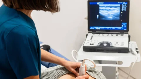
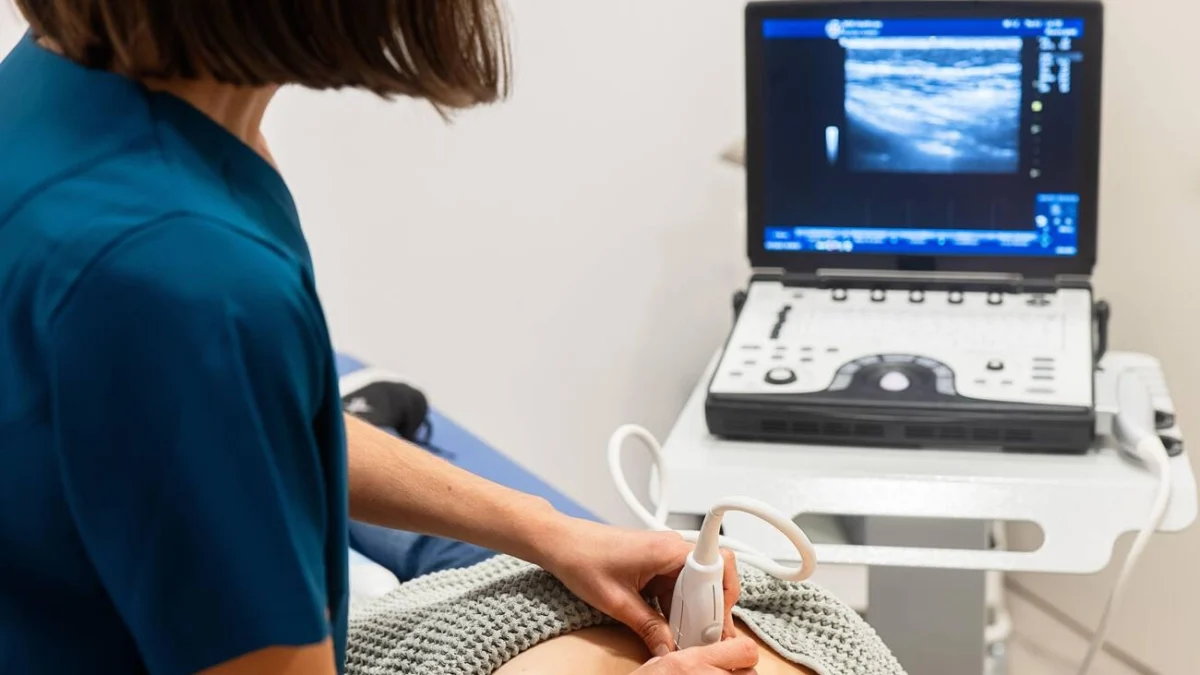
For accurate diagnosis, modern medicine has progressed far beyond standard Liver Function Tests (LFTs). More than 50% of individuals with biopsy-proven steatosis possess completely normal ALT and AST blood levels! Today’s gold-standard non-invasive evaluation is Transient Elastography (FibroScan). Using low-frequency shear waves, FibroScan simultaneously measures two vital parameters: the Controlled Attenuation Parameter (CAP score), which grades fat volume from S1 to S3, and Liver Stiffness Measurement (LSM in kilopascals), which accurately detects fibrotic scarring from F0 to F4 without requiring a painful needle biopsy [1].
The 90-Day Reversal Blueprint: Nutrition and Exercise Protocols
The remarkable virtue of human hepatic biology is its extraordinary capacity for regeneration. Unlike the central nervous system or myocardium, the liver can completely purge accumulated fat and regenerate healthy functional tissue within 90 days when physiological roadblocks are removed [3]:
1. Eliminate Liquid Fructose and Refined Sugars: Banish commercial sodas, sweetened teas, packaged fruit juices, and hidden high-fructose corn syrup. Removing the primary biochemical substrate for De Novo Lipogenesis halts fat synthesis within days [2].
2. Time-Restricted Eating (16:8 Fasting): Limiting food consumption to an 8-hour daily window allows circulating insulin levels to plummet overnight. Once insulin falls to baseline, the liver activates hepatic lipase enzymes, switching from fat storage to active intrahepatic fat oxidation [3].
3. Post-Meal Physical Movement: Engaging in light, consistent physical activity, such as walking after meals and postprandial glucose disposal, activates GLUT4 transporters in skeletal muscle without requiring insulin, diverting circulating sugars directly into working muscle cells before the liver can convert them into fat [2], [3].
4. Prioritize Choline, Polyphenols, and Omega-3s: Ensure adequate dietary choline (from pastured eggs and cruciferous vegetables) to facilitate VLDL fat export, alongside extra virgin olive oil, green tea, and wild fatty fish to neutralize oxidative lipotoxicity [1], [4].
The Twin Cycle Hypothesis: Restoring Long-Term Metabolic Freedom
Pioneering clinical investigations by Professor Roy Taylor at Newcastle University established the Twin Cycle Hypothesis of metabolic disease. Taylor demonstrated that Type 2 diabetes and MASLD are interconnected loops of ectopic fat accumulation: fat in the liver spills over into the pancreas, paralyzing insulin-secreting beta cells [3].
Critically, Taylor’s clinical trials proved that losing just 5% to 10% of total body weight (roughly a single gram of fat cleared from the liver and pancreas) completely reverses hepatic insulin resistance, normalizes fasting blood glucose, and restores pristine liver architecture. A fatty liver is not a permanent life sentence; it is a reversible metabolic consequence of modern dietary excess [1], [3].
In conclusion, the quiet warning of a fatty liver diagnosis should be received not with despair, but as a priceless physiological wake-up call. By understanding the metabolic biochemistry of De Novo Lipogenesis and implementing disciplined nutritional and physical habits, you hold the power to completely clear liver fat within 90 days, reclaiming your health, energy, and longevity from the inside out [1], [3].
Frequently Asked Questions
Common reader questions about this study
Can non-drinkers develop severe fatty liver disease?
Yes. Non-alcoholic fatty liver, now medically termed MASLD, is primarily driven by excess dietary refined carbohydrates, high-fructose corn syrup, and systemic insulin resistance, which mimic the exact same biochemical lipogenic pathways as chronic alcohol consumption.
Is Grade 1 Fatty Liver dangerous or curable?
Grade 1 steatosis is the earliest stage, where fat droplets accumulate in 5% to 33% of hepatocytes without active inflammation or scar tissue. It is 100% reversible within 8 to 12 weeks through dietary carbohydrate restriction and routine physical exercise.
What is the most accurate diagnostic test for liver fat and scarring?
While conventional abdominal ultrasound detects fat accumulation once it exceeds 20%, transient elastography (FibroScan) provides non-invasive, quantitative measurements of both fat steatosis (CAP score in dB/m) and tissue stiffness or fibrosis (kPa score).
What is the connection between fatty liver and Type 2 diabetes?
They are two clinical manifestations of the same underlying metabolic syndrome. Ectopic fat accumulation in the liver induces hepatic insulin resistance, causing uncontrolled nocturnal gluconeogenesis that spikes fasting blood glucose levels and precipitates Type 2 diabetes.
References
- ↩ Rinella ME, et al. A multisociety Delphi consensus statement on new fatty liver disease nomenclature. Ann Hepatol. 2023;29(1):101133
- ↩ Petersen MC, Shulman GI. Mechanisms of Insulin Action and Insulin Resistance. Physiol Rev. 2018;98(4):2133-2223
- ↩ Taylor R, et al. Type 2 diabetes and remission: the role of the twin cycles. Lancet Diabetes Endocrinol. 2018;6(6):422-424
- ↩ Anjana RM, et al. Metabolic non-communicable disease health report in India: ICMR-INDIAB. Lancet Diabetes Endocrinol. 2023;11(7):474-489


No comments yet. Be the first to join the conversation.