


Key Findings
- High insulin sensitivity enables cells to clear circulating glucose with minimal pancreatic insulin secretion, preventing hyperinsulinemia and visceral fat deposition.
- Skeletal muscle accounts for over 80% of postprandial glucose uptake, making muscular contractions the most potent non-pharmacological stimulus for GLUT4 translocation.
- A single night of partial sleep deprivation (4 hours) reduces acute peripheral insulin sensitivity by 25% to 30% via sympathetic overdrive and cortisol surges.
- Dietary strategies emphasizing soluble prebiotic fibers and resistant starches generate short-chain fatty acids (SCFAs) that improve hepatic insulin signaling.
Have you ever paused to consider how the energy from the food you eat actually journeys into the trillions of living cells that animate your body? Whether you consume a warm breakfast or a balanced lunch, dietary carbohydrates are systematically broken down by digestive enzymes into a simple six-carbon sugar called glucose. As glucose streams into systemic circulation, it cannot simply diffuse across cellular lipid membranes on its own. It requires an exquisitely precise biological key to unlock cellular gates. That master biochemical key is insulin—a peptide hormone synthesized and secreted by the beta cells of the endocrine pancreas [1].
Imagine the exterior of a human cell as a reinforced door fitted with a precision lock. When the lock is well-oiled and perfectly maintained, inserting the key turns the deadbolt smoothly with zero resistance, swinging the door wide open. In metabolic biology, when muscle and liver cells recognize minuscule quantities of insulin and immediately mobilize glucose transport channels, that state of effortless efficiency is termed insulin sensitivity. The higher your insulin sensitivity, the more agile, youthful, and resilient your whole-body metabolism remains. Conversely, when that lock becomes gummed up with ectopic fat metabolites, the key refuses to turn—forcing the pancreas to forge dozens of identical keys (hyperinsulinemia) just to push the door open. Understanding this balance is the key to reversing insulin resistance, avoiding fatty liver disease, and sustaining lifelong metabolic vitality [1], [4].
- 80%+
- Muscle Glucose Clearance Skeletal muscle absorbs the vast majority of postprandial glucose
- < 1.0
- Optimal HOMA-IR Score Clinical biomarker indicating superior cellular insulin sensitivity
- 48 Hours
- Exercise Legacy Effect Duration of enhanced muscle glucose uptake following a workout
- 25-30%
- Sensitivity Drop Acute insulin desensitization caused by a single night of sleep loss
Molecular Mechanics: How Insulin Unlocks the Cellular Door
To appreciate how insulin sensitivity protects human health, we must zoom down to the nanoscopic architecture of the cellular membrane. Every skeletal muscle fiber and hepatocyte is studded with specialized transmembrane proteins known as insulin receptors. When postprandial blood glucose concentrations rise, pancreatic beta cells release pulses of insulin that travel through the bloodstream and dock precisely onto the alpha-subunits of these receptors [1].
This binding event triggers autophosphorylation of the receptor’s intracellular beta-subunits, unleashing a swift biochemical signaling cascade known as the PI3K-Akt signaling pathway. Specifically, an enzyme called phosphoinositide 3-kinase (PI3K) activates a master kinase named Akt2. Phosphorylated Akt2 immediately neutralizes an inhibitory protein called AS160 (Akt substrate of 160 kDa), which normally keeps specialized transport vesicles anchored inside the cell interior. Once AS160 is inhibited, intracellular vesicles carrying GLUT4 (Glucose Transporter Type 4) proteins surge toward the cell periphery, fusing seamlessly into the outer plasma membrane [2].
Once embedded in the cellular surface, GLUT4 transporters act as open doorways, allowing blood glucose to rush down its concentration gradient into the intracellular fluid. Inside muscle cells, mitochondria oxidize this glucose into adenosine triphosphate (ATP) for immediate muscular contraction, while surplus glucose is linked into long branched chains of glycogen for emergency energy reserves. Once circulating glucose returns to baseline, the cell retrieves GLUT4 channels back into storage vesicles, resetting the system. When an individual possesses high insulin sensitivity, this entire molecular sequence executes flawlessly with minimal insulin secretion, preventing arterial inflammation and pancreatic exhaustion [1], [2].
From an evolutionary perspective, our ancestral hunter-gatherer biology evolved under conditions of chronic food scarcity punctuated by brief feasts following a successful hunt. Superior insulin sensitivity ensured that rare nutritional carbohydrates were instantly shunted into skeletal muscles rather than lost. However, in our modern environment of continuous hyper-caloric abundance and physical stillness, cellular glycogen tanks remain chronically full, causing the signaling cascade to break down and inducing cellular resistance [3], [4].
The Cellular Cascade of Insulin Action
Systemic Repercussions: What Peak Sensitivity Feels Like
When your body operates with optimal insulin sensitivity, you experience what metabolic physiologists describe as metabolic flexibility. A metabolically flexible human functions like an advanced hybrid automobile: when carbohydrates are consumed, the body seamlessly burns glucose; when fasting between meals or sleeping overnight, it effortlessly pivots to oxidizing stored body fat for fuel [1].
This metabolic agility prevents the roller-coaster crashes of reactive hypoglycemia that plague desk workers. Furthermore, high insulin sensitivity dramatically improves cardiovascular hemodynamics. Insulin stimulates endothelial cells lining blood vessel walls to produce nitric oxide (NO)—a potent gaseous vasodilator that relaxes vascular smooth muscle, reduces peripheral vascular resistance, and maintains healthy baseline blood pressure [1], [4].
In the liver, sensitive hepatocytes efficiently suppress unnecessary gluconeogenesis (the endogenous synthesis of new glucose from amino acids) once you have eaten. Because the liver is not overwhelmed by high circulating insulin and substrate overflow, de novo lipogenesis remains turned off, completely preventing triglyceride deposition and hepatic steatosis. Systemic inflammatory biomarkers such as high-sensitivity C-reactive protein (hs-CRP) and interleukin-6 remain suppressed, protecting joints, microvascular beds, and skin integrity [1], [3].
The Brain Connection: Preventing Type 3 Diabetes
While historically viewed as a peripheral hormone confined to liver, fat, and muscle biology, neuroscientists now recognize insulin as a critical master regulator of cerebral bioenergetics. The human brain accounts for barely 2% of total body mass yet consumes over 20% of whole-body glucose energy. Within the central nervous system, dense populations of insulin receptors are concentrated in the hippocampus—the neuroanatomical epicenter of memory consolidation, spatial navigation, and synaptic plasticity [1].
When hippocampal neurons maintain sharp insulin sensitivity, insulin signaling upregulates Brain-Derived Neurotrophic Factor (BDNF), fortifying synaptic connections and stimulating adult neurogenesis. However, when systemic insulin resistance spills over into the central nervous system, cerebral glucose hypometabolism ensues. Starved of energetic substrates, hippocampal neurons suffer synaptic degeneration and accelerated apoptosis [1].
Furthermore, cerebral insulin resistance directly accelerates the pathology of Alzheimer’s disease. The primary enzyme responsible for degrading circulating insulin in the brain is Insulin-Degrading Enzyme (IDE). Crucially, IDE is also the principal enzyme tasked with clearing neurotoxic amyloid-beta plaques. When chronic hyperinsulinemia floods the cerebral spinal fluid with excess insulin, IDE is monopolized by breaking down insulin, leaving amyloid-beta peptides free to aggregate into neurofibrillary tangles. Because of this shared pathophysiological etiology, progressive neurodegenerative dementia is increasingly classified in clinical research as Type 3 Diabetes [1].
| Diagnostic Biomarker | High Insulin Sensitivity | Early Insulin Resistance | Advanced Type 2 Diabetes |
|---|---|---|---|
| Fasting Blood Glucose | 70 to 89 mg/dL | 90 to 99 mg/dL | 126 mg/dL or higher |
| Fasting Serum Insulin | 2 to 5 uIU/mL | 8 to 15 uIU/mL | > 20 uIU/mL (or beta cell exhaustion) |
| HOMA-IR Score | Less than 1.0 | 1.5 to 2.5 | Greater than 3.0 |
| Triglyceride-to-HDL Ratio | Less than 1.5 | 2.0 to 3.5 | Greater than 4.0 |
| Metabolic Flexibility | Rapid switch from carb to fat burn | Sluggish fat oxidation | Complete reliance on glucose, constant hunger |
Data adapted from Yale Endocrinology Clamp Protocols and ICMR-INDIAB Studies
Five Clinically Proven Pathways to Boost Insulin Sensitivity
Reclaiming optimal insulin sensitivity does not require restrictive starvation diets or exotic pharmaceutical compounds. By strategically leveraging skeletal muscle physiology, chronobiology, and nutritional biochemistry, you can systematically reboot your cellular insulin receptors through five evidence-based interventions [1], [2]:
1. Build Skeletal Muscle Mass (Resistance Training): Skeletal muscle is the primary metabolic sink of the human body, disposing of 80% or more of circulating glucose following a meal. Engaging in progressive resistance training—such as squats, deadlifts, push-ups, and pull-ups—depletes intramyocellular glycogen stores. Furthermore, vigorous muscular contraction activates AMP-activated protein kinase (AMPK), which drives GLUT4 transporters to the cell membrane entirely independent of insulin. Each workout enhances muscle insulin sensitivity for up to 48 hours [2].
2. Implement Post-Meal Walking Breaks: Instead of collapsing on a sofa after lunch or dinner, commit to a 10-to-15-minute gentle walk. Contracting large leg muscles immediately after eating blunts postprandial glucose excursions by up to 30%, clearing glucose directly from the bloodstream before the pancreas is forced to flood the body with high insulin spikes. For office professionals, integrating walking after meals to blunt glucose spikes is the single most accessible habit for metabolic defense [1], [4].
3. Protect Deep Non-REM Sleep: Sleep is not passive rest; it is active metabolic recalibration. Landmark sleep laboratory trials demonstrate that subjecting healthy young adults to just one single night of 4-hour sleep restriction reduces peripheral insulin sensitivity by 25% to 30%. Sleep deprivation triggers nocturnal sympathetic nervous system hyperactivity, surges morning cortisol, and blunts growth hormone pulsatility. Prioritize 7 to 8 hours of uninterrupted nocturnal rest in a cool, dark environment to keep cellular receptors responsive [1].
4. Prioritize Fermentable Fibers and Resistant Starches: Dietary fiber is the essential fuel for your colonic microbiome. When gut bacteria ferment soluble prebiotic fibers (found in oats, legumes, flaxseeds, and cooked-and-cooled potatoes), they generate bioactive short-chain fatty acids (SCFAs), predominantly acetate, propionate, and butyrate. Butyrate repairs gut barrier integrity, preventing inflammatory bacterial endotoxins (LPS) from leaking into the portal vein, while propionate directly downregulates hepatic glucose output, dramatically improving liver insulin sensitivity [1], [3].
5. Adopt an Early Time-Restricted Feeding Window: Aligning food intake with your body’s circadian clock optimizes hormone dynamics. Human insulin sensitivity peaks in the morning and early afternoon, steadily deteriorating as night approaches due to the natural nocturnal surge of melatonin. Consuming all daily meals within an 8-to-10-hour daytime window (e.g., 8:00 AM to 6:00 PM) while keeping evenings fasted allows baseline insulin levels to fall, reactivating hormone-sensitive lipase (HSL) and clearing ectopic lipid accumulation from muscle and liver tissue [1], [4].
Diagnostic Testing: How to Measure Your Sensitivity
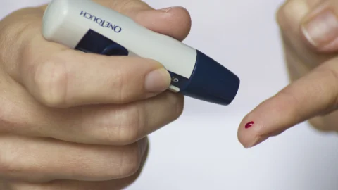
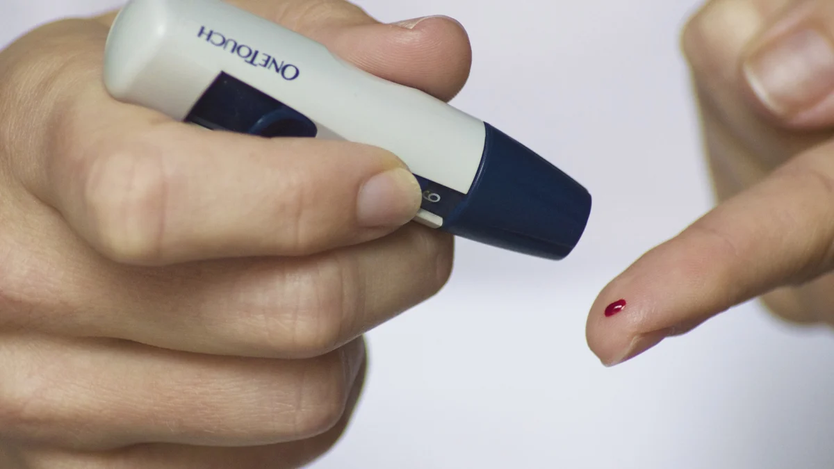
Standard fasting blood sugar tests often fail to detect metabolic dysfunction until late in the disease trajectory; a person can maintain normal blood sugar for over a decade simply because their pancreas is pumping out four times the normal amount of insulin. To evaluate your true cellular sensitivity, request these two critical lab tests [1], [4]:
Fasting Serum Insulin: While standard fasting glucose measures how much sugar is in your blood, fasting insulin reveals how hard your pancreas is working to maintain that number. An optimal fasting insulin level falls between 2 and 5 uIU/mL. A fasting insulin level above 10 uIU/mL—even with a normal fasting blood sugar of 85 mg/dL—is a definitive indicator of early underlying insulin resistance.
The HOMA-IR Calculation: Using your fasting glucose (in mg/dL) and fasting insulin (in uIU/mL), calculate your Homeostatic Model Assessment of Insulin Resistance: HOMA-IR = (Fasting Glucose × Fasting Insulin) / 405. A score below 1.0 represents peak insulin sensitivity. Scores between 1.0 and 1.9 indicate emerging resistance, while values exceeding 2.0 indicate significant metabolic resistance requiring immediate lifestyle intervention [4].
Circadian Biology: Melatonin, Chrono-Nutrition and Meal Timing
One of the most frequently overlooked regulators of insulin sensitivity is your biological circadian clock. In human chronobiology, metabolic efficiency fluctuates predictably across a 24-hour cycle. Peripheral insulin sensitivity is at its biological zenith during morning and early afternoon daylight hours, steadily diminishing as dusk approaches [1].
This diurnal variation is directly orchestrated by the pineal hormone melatonin. As darkness falls, melatonin secretion surges to prepare the brain for sleep. However, pancreatic beta cells and hepatocytes express dense melatonin receptors (MT1 and MT2). Melatonin binding to beta cells acts as an evolutionary brake, intentionally suppressing insulin secretion and blunting glucose clearance so that glucose remains available for cerebral maintenance during overnight fasting. When individuals consume heavy, carbohydrate-laden dinners late at night, they are dumping glucose into a biological system that has actively switched off its insulin machinery—triggering prolonged nocturnal hyperglycemia, systemic inflammation, and impaired morning insulin sensitivity [1], [4].
Frequently Asked Questions
Common reader questions about this study
What is the physiological difference between insulin sensitivity and insulin resistance?
Insulin sensitivity measures how effectively cells open glucose gates in response to small amounts of insulin. In contrast, insulin resistance occurs when cellular receptors become desensitized, forcing the pancreas to hyper-secrete insulin to manage the same carbohydrate load.
How does physical exercise improve insulin sensitivity without needing insulin?
Muscular contraction stimulates AMP-activated protein kinase (AMPK), which mechanically drives GLUT4 glucose transporter vesicles directly to the cell membrane, clearing glucose entirely independent of insulin signaling pathways.
What is considered an ideal HOMA-IR score for metabolic health?
A Homeostatic Model Assessment for Insulin Resistance (HOMA-IR) score below 1.0 indicates optimal insulin sensitivity. Values between 1.0 and 1.9 indicate early insulin resistance, while scores above 2.0 reflect established metabolic resistance.
How long do the insulin-sensitizing effects of a single workout last?
Following a session of moderate-to-vigorous resistance or endurance training, skeletal muscle insulin sensitivity remains significantly elevated for 24 to 48 hours as glycogen-depleted muscles absorb circulating glucose to rebuild fuel reserves.
References
- ↩ Petersen MC, Shulman GI. Mechanisms of Insulin Action and Insulin Resistance. Physiol Rev. 2018;98(4):2133-2223
- ↩ Richter EA, Hargreaves M. Exercise, GLUT4, and skeletal muscle glucose uptake. Physiol Rev. 2013;93(3):993-1017
- ↩ Anjana RM, et al. Metabolic non-communicable disease health report in India: ICMR-INDIAB. Lancet Diabetes Endocrinol. 2023;11(7):474-489
- ↩ DeFronzo RA. From the Triumvirate to the Ominous Octet: A New Paradigm for Type 2 Diabetes. Diabetes. 2009;58(4):773-795

No comments yet. Be the first to join the conversation.