




Key Findings
- Sudden death in young athletes under 35 is rarely caused by coronary artery plaque blockages; silent structural anomalies like hypertrophic cardiomyopathy and primary electrical channelopathies trigger more than 80 percent of fatal events.
- Data from 10.9 million marathon and half-marathon runners reveal an overall sudden cardiac arrest incidence of 0.54 per 100,000, with men accounting for 86 percent of all race-associated arrests.
- Bystander cardiopulmonary resuscitation (CPR) combined with automated external defibrillator (AED) delivery within 3 to 5 minutes elevates survival odds past 70 percent.
A tenth-grade student finishes an afternoon physical education session on a sunlit school track, walks back to his homeroom, and collapses forward onto his desk. Across town during an annual city marathon, a 24-year-old amateur runner collapses 400 meters before the finish line, completely unresponsive and without a palpable carotid pulse. These catastrophic events recur with disturbing regularity across global headlines, leaving parents, coaches, and sports physicians grasping for answers. The victims are visibly fit, carry no clinical history of hypertension or smoking, and frequently represent the most athletic individuals in their peer groups. Why would a young, well-conditioned body abruptly surrender to fatal circulatory collapse during or immediately following exercise?
Cardiovascular medicine does not view these tragic episodes as inexplicable accidents. Forensic pathology demonstrates that strenuous physical exertion serves as an unsparing hemodynamic stress test, unmasking quiescent anatomical malformations and electrical vulnerabilities that remain entirely undetectable during quiet daily life. Landmark clinical statements from the American Academy of Pediatrics (AAP) confirm this distinction [1]. Large-scale registry data in the New England Journal of Medicine (NEJM) show that the biological drivers of sudden death in youth differ fundamentally from older adults [2]. Deconstructing these mechanisms provides the evidence base required to protect young participants on playing fields and racecourses worldwide.
- 10.9 Million
- Runners Monitored NEJM RACER 10.5-year multi-race cohort
- 0.54 / 100,000
- Overall Arrest Rate Rare but highly lethal clinical event
- 3.70x Risk
- Unaccustomed Exertion ICMR multi-center matched case-control finding
- 86%
- Male Predominance Observed across distance running cardiac arrests
The Shock of Sudden Cardiac Arrest in Young Athletes
Public perception overwhelmingly links heart failure and sudden death to advanced age, sedentary lifestyles, tobacco dependency, and calcified arteries. When an adolescent footballer or collegiate middle-distance runner drops motionless to the turf, surrounding spectators reflexively suspect acute dehydration, heat exhaustion, severe hypoglycemia, or a primary neurological seizure. This cognitive bias and the resulting diagnostic hesitation frequently consume the few minutes during which the patient’s brain can still be salvaged. Sudden cardiac arrest (SCA) in athletes under 35 strikes 1 in 40,000 to 1 in 80,000 participants annually [1]. Exact event rates depend heavily on sport intensity, competition level, and gender.
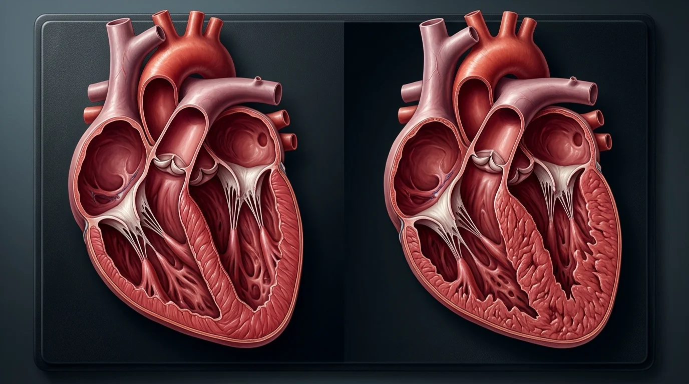
Sustained athletic conditioning triggers profound physiological remodeling within the human heart. Long-term volume and pressure overloads lead to increased left ventricular chamber dimensions, enhanced myocardial compliance, and resting vagal bradycardia, a benign condition recognized clinically as the athlete heart. Under ordinary circumstances, these structural adaptations optimize stroke volume and cardiac reserve. However, rigorous training can unmask an occult genetic or structural defect. A sudden surge in circulating adrenaline and myocardial oxygen demand transforms the heart into an erratic generator of lethal arrhythmias.
Heart Attack vs Sudden Cardiac Arrest: The Core Clinical Difference
Mainstream news reports almost universally describe athletic deaths as massive heart attacks. In clinical cardiology, a heart attack (myocardial infarction) and sudden cardiac arrest represent entirely different pathophysiological mechanisms. A myocardial infarction is fundamentally a circulatory plumbing failure. Rupture of a fragile, lipid-laden atheromatous plaque within the intimal layer of a coronary artery triggers acute thrombosis, cutting off oxygenated blood supply to downstream myocardium. The patient typically remains conscious, experiencing progressive, crushing retrosternal chest pain, diaphoresis, and shortness of breath while myocardial tissue undergoes ischemic necrosis over several hours.
Sudden cardiac arrest, by contrast, is an acute electrical catastrophe. The heart’s organized electromechanical conduction pathway degenerates abruptly into chaotic, uncoordinated depolarizations, most commonly ventricular fibrillation (VF). Under ventricular fibrillation, the ventricles flutter at rates exceeding 300 beats per minute, rendering mechanical stroke volume nonexistent. Forward arterial blood pressure collapses to zero in a fraction of a second. Deprived of oxygenated arterial flow, cerebral cortical neurons lose function within 6 to 10 seconds, causing the athlete to collapse into unresponsiveness without time to brace for impact. The comparative breakdown below outlines these critical clinical differences.
| Feature | Heart Attack (Myocardial Infarction) | Immediate ThreatSudden Cardiac Arrest (SCA) |
|---|---|---|
| Primary Pathology | Coronary artery thrombotic occlusion (Plumbing failure) | Chaotic ventricular tachyarrhythmia (Electrical failure) |
| Myocardial Motion | Heart continues beating; focal ischemia develops | Mechanical pumping halts; ventricles quiver helplessly |
| Consciousness | Conscious; experiences chest pressure and dyspnea | Immediate loss of consciousness within 5 to 10 seconds |
| Breathing Pattern | Labored breathing persists | Respiration ceases; agonal gasps may occur briefly |
| Primary Intervention | Emergency coronary catheterization and thrombolysis | Immediate bystander CPR and rapid AED defibrillation |
Source: American Heart Association (AHA) and American College of Cardiology Guidelines
While an acute myocardial infarction allows an operative emergency window spanning several hours to achieve percutaneous coronary intervention (PCI), sudden cardiac arrest permits a therapeutic rescue window measured strictly in minutes. After four minutes of circulatory standstill, irreversible anoxic encephalopathy begins; after ten minutes without resuscitation, functional neurological survival is vanishingly rare. Just as modern surgical teams develop artificial solutions like the world first heart made from titanium, saving an athlete requires fast on-site action. Resuscitation must happen before advanced hospital transport arrives.
Five Hidden Cardiovascular Defects Behind Youth Fatalities
Forensic autopsy registries in young athletes demonstrate that non-traumatic sudden cardiac death stems overwhelmingly from five distinct congenital, structural, and electrophysiological substrates [1]. These silent conditions remain asymptomatic throughout childhood, evading routine pediatric checkups until maximum physical exertion acts as a lethal trigger.
1. Hypertrophic Cardiomyopathy (HCM): HCM is the leading identified cause of non-traumatic sudden athletic death in western registries. It is an inherited genetic condition affecting roughly 1 in 500 individuals. Point mutations in sarcomeric protein genes (notably MYH7 and MYBPC3) trigger progressive, asymmetrical thickening of the left ventricular myocardium, most commonly involving the basal interventricular septum. Microscopic examination reveals profound myocyte disarray, chaotic branching, and dense interstitial fibrosis. During heavy sprinting, dynamic left ventricular outflow tract (LVOT) obstruction combines with severe subendocardial ischemia to generate lethal re-entrant ventricular tachycardia.
2. Anomalous Aortic Origin of a Coronary Artery (AAOCA): In this congenital vascular flaw, a coronary artery arises from the wrong aortic sinus. Most dangerously, the left main coronary artery emerges anomalously from the right sinus of Valsalva. The anomalous artery must take an acute, angulated course traversing directly between the aortic root and the main pulmonary artery. During intense athletics, expansion of both great vessels under elevated systolic pressures compresses the slit-like coronary orifice and intramural arterial segment. This mechanical pinch throttles left ventricular blood supply at peak demand, precipitating acute transmural ischemia and sudden ventricular collapse.
3. Primary Ion Channelopathies: Certain collapsed athletes show no structural abnormalities whatsoever on gross post-mortem dissection or pre-participation echocardiography. These patients harbor inherited channelopathies, which are genetic mutations in myocardial transmembrane ion channels governing sodium, potassium, or calcium conductances. Long QT Syndrome (LQTS) delays ventricular repolarization, making the heart susceptible to torsades de pointes. Catecholaminergic Polymorphic Ventricular Tachycardia (CPVT), linked to mutations in the ryanodine receptor gene (RYR2), causes bidirectional ventricular tachycardia triggered exclusively by high adrenergic tone during intense athletic sprints or severe emotional fright.
4. Acute and Subacute Myocarditis: Inflammation of the myocardium secondary to common cardiotropic viral infections (including enteroviruses, parvovirus B19, adenoviruses, and post-viral systemic syndromes) causes focal myocyte necrosis and patchy replacement fibrosis. While constitutional viral symptoms like fever and myalgia often resolve within days, microscopic myocardial inflammation and electric instability linger for weeks. Vigorous exercise undertaken before complete tissue healing provokes malignant ventricular arrhythmias. Broader metabolic research, such as studies evaluating circadian rhythms in eating after 9pm and metabolic health, highlights this balance. Cellular recovery and restorative rest dictate tissue resilience across all vital organs.
5. Commotio Cordis: In commotio cordis, an entirely healthy heart with no structural or electrical defects is arrested by blunt trauma. It occurs when a dense projectile, such as a baseball, cricket ball, or hockey puck, strikes the anterior precordium directly over the left ventricle. Timing is critically vulnerable. If the impact hits during a 15 to 30 millisecond window on the electrocardiographic T-wave upslope, mechanical deformation triggers stretch-sensitive ion channels, inducing immediate ventricular fibrillation.
Hemodynamic Stress and Electrical Collapse During Intense Exertion
During quiet rest, the healthy heart beats 60 to 80 times per minute, circulating approximately 5 liters of blood through the systemic vasculature each minute. During competitive distance racing or high-intensity interval training, working skeletal muscles require immense volumes of oxygenated hemoglobin and rapid lactate clearance. To satisfy this demand, the cardiovascular system increases total cardiac output according to the standard hemodynamic formulation:
During peak exertion, an athlete’s heart rate climbs toward 180 to 200 beats per minute, while ventricular stroke volume nearly doubles. Total cardiac output can escalate dramatically from 5 liters per minute up to 30 or 35 liters per minute in elite endurance competitors. This monumental mechanical output requires an exponential expansion in coronary blood flow to nourish the vigorously contracting myocardium. In a normal cardiovascular system, metabolic autoregulation dilates coronary epicardial arteries and intramyocardial arterioles, lowering vascular resistance to deliver adequate oxygen.
In an athlete harboring an occult structural or electrical anomaly, this compensatory physiology collapses. Severe tachycardia substantially truncates the diastolic filling interval, which is the only phase of the cardiac cycle when coronary perfusion occurs. In hypertrophied muscle, elevated intracavitary pressures and thickened muscle bundles mechanically compress intramural vessels. When oxygen delivery drops below critical metabolic thresholds, subendocardial myocytes rapidly accumulate lactic acid and lose intracellular potassium. This regional heterogeneity in action potential duration destabilizes myocardial electrical conduction, setting off the fatal sequence mapped out below.
Physiological Cascade of Exercise Induced Sudden Cardiac Arrest
- Stage 1
Sympathetic Adrenergic Surge
Intense athletic exertion elevates circulating epinephrine and norepinephrine, pushing heart rates past 180 beats per minute. - Stage 2
Subendocardial Ischemic Strain
Shortened diastole and compressed anomalous vessels severely compromise coronary perfusion to the thickened myocardium. - Stage 3
Electrophysiological Instability
Hypoxic myocytes develop altered resting membrane potentials and fragmented conduction, generating re-entrant wavefronts. - Stage 4
Ventricular Fibrillation
Organized mechanical ventricular contractions collapse into chaotic electrical quivering; forward cardiac output drops to zero. - Stage 5
Cerebral Anoxia and Collapse
Within 6 to 10 seconds, oxygen deprivation causes abrupt unresponsiveness, requiring immediate chest compressions.
Methods: How the NEJM and ICMR Studies Were Conducted
To establish the true incidence, clinical characteristics, and risk factors underlying exercise-associated sudden death, medical researchers created large-scale, prospective and retrospective population databases. In the United States, the Race Associated Cardiac Arrest Event Registry (RACER) study, led by Dr. Jonathan H. Kim and Dr. Aaron L. Baggish and published in the New England Journal of Medicine, monitored all participants in certified US marathons and half-marathons over a 10.5-year period from January 1, 2000, through May 31, 2010 [2]. The investigators analyzed on-site medical logs, EMS run sheets, survivor hospital records, and medical examiner autopsy reports across 10,905,015 runner participations.
In India, public concern over sudden unexplained deaths in young adults prompted a landmark scientific investigation. The Indian Council of Medical Research (ICMR) and NIE launched an extensive multi-center matched case-control study [3]. Conducted across 47 tertiary hospitals in 19 Indian states, the study evaluated 729 unexplained sudden deaths between October 2021 and March 2023. All participants were aged 18 to 45 and visibly healthy within 24 hours of collapse. Investigators matched each case with 4 healthy neighborhood controls (2,916 total controls) of identical age and sex. This allowed clinicians to evaluate vaccination records, family pedigrees, and recent vigorous exercise patterns.
Key Epidemiological Findings: Marathon Registries and Youth Risk Factors
The RACER study provided clear, objective data defining the risk landscape of distance running. Over the 10.5-year observation window, 59 runners suffered cardiac arrest across 10.9 million participations, yielding an overall incidence rate of 0.54 per 100,000 runners. The risk was concentrated in full marathons (1.01 per 100,000) compared to half-marathons (0.27 per 100,000). A profound gender disparity emerged: men accounted for 86 percent of all cardiac arrest events, experiencing an event rate of 0.90 per 100,000 compared to just 0.16 per 100,000 for women.
As illustrated in Chart 1, male full marathon participants experienced the highest event rate at 2.03 per 100,000 runners during the study decade. Clinical and forensic follow-up revealed that older athletes aged 35 and above overwhelmingly succumbed to atherosclerotic coronary artery disease, whereas younger runners died from hypertrophic cardiomyopathy or congenital coronary anomalies. Just as global health analyses exploring global obesity and metabolic disease reveal distinct age-dependent epidemiological shifts, the physiological drivers of athletic cardiac arrest split sharply at the 35-year age boundary.
As detailed in Table 1, the pathophysiological drivers behind youth sudden cardiac arrest stand in sharp contrast to those observed in older master athletes. While master athletes suffer from intimal plaque rupture and luminal occlusion, younger athletes harbor inherited structural, histological, and electrical abnormalities that remain clinically silent until stressed by extreme exercise.
| Clinical Parameter | Young Athletes (<35 Years) | Master Athletes (≥35 Years) |
|---|---|---|
| Primary Etiology | Hypertrophic cardiomyopathy, anomalous coronary arteries, channelopathies | Atherosclerotic coronary artery disease, plaque rupture |
| Pathological Substrate | Congenital architectural defects, cellular disarray, genetic ion mutations | Luminal stenosis, calcified fibroatheroma, intimal inflammation |
| Primary Screening Test | 12-lead ECG, transthoracic echocardiogram, genetic panels | Exercise treadmill test (TMT), CT coronary calcium scoring, angiogram |
| Exercise Trigger | Sympathetic surge initiates re-entrant ventricular tachyarrhythmias | High myocardial workload provokes plaque rupture and coronary thrombosis |
Source: American Academy of Pediatrics (AAP) and New England Journal of Medicine (NEJM) data
The ICMR investigation in India yielded equally consequential findings regarding young adult mortality. The study conclusively demonstrated that COVID-19 vaccination did not increase the risk of sudden unexplained death; in fact, vaccinated individuals exhibited reduced odds of sudden death (adjusted matched odds ratio [aOR] 0.51). Conversely, the study identified that engaging in vigorous, unaccustomed physical activity within 48 hours of collapse increased the risk of sudden unexplained death by 3.70 times. Having a family history of premature sudden death raised the risk by 2.53 times, while heavy binge drinking increased the risk by 5.29 times. These findings reinforce that intense physical exertion without gradual conditioning poses severe hazards for individuals with unmasked cardiovascular vulnerabilities.
Early Warning Flags Parents and Athletic Coaches Must Recognize
Sudden cardiac arrest seems abrupt, but warning signs often precede collapse. Retrospective data from the American Academy of Pediatrics show that up to 50 percent of young victims experience antecedent symptoms weeks earlier [1]. Unfortunately, athletes frequently conceal these episodes, while parents and coaches mistakenly attribute them to fatigue, dehydration, or warm weather.
Clinicians, coaches, and school administrators must maintain heightened vigilance for four cardinal cardiovascular red flags:
1. Exertional Syncope or Pre-Syncope: Loss of consciousness occurring during active exercise is an ominous clinical sign that should never be dismissed as benign vasovagal syncope. Fainting that occurs mid-stride or during exertion indicates sudden, profound cerebral hypoperfusion, often caused by dynamic left ventricular outflow tract obstruction or self-terminating ventricular tachyarrhythmias.
2. Disproportionate Exertional Chest Pressure: Unexplained retrosternal tightness, constriction, or radiating pain during athletic drills signals severe myocardial ischemia, frequently indicating an anomalous coronary artery compressed between the great arteries.
3. Unprovoked Rapid Palpitations: Sensations of sudden, racing, or skipped beats during rest or minimal exertion suggest underlying supraventricular or ventricular arrhythmias, including latent channelopathies like long QT syndrome.
4. Family History of Premature Sudden Death: Any unexplained sudden death or drowning in a relative under age 50 requires attention. First-degree relatives must undergo a 12-lead ECG and echocardiogram before entering competitive sports.
Study Limitations: What the Registries Cannot Tell Us
While the RACER registry and ICMR investigations provide invaluable epidemiological insights, critical methodological boundaries remain. First, retrospective race registries depend on paramedic event logs and post-mortem coroner examinations that vary across municipal jurisdictions. Incomplete molecular autopsy panels in historical cases mean that certain subtle ion channelopathies may have been misclassified as unexplained or idiopathic arrests.
Second, population-level matched case-control studies evaluate statistical associations rather than direct biological causality. Unaccustomed vigorous exercise showed a 3.7-fold increase in sudden death odds. Even so, researchers cannot track every variable, such as asymptomatic viral myocarditis, acute electrolyte deficits, or stimulant supplement abuse. Future prospective athletic registries utilizing widespread pre-season genetic sequencing and wearable ECG telemetry will be required to establish definitive causal pathways.
What Happens Next: Preventive Action and Emergency Lifesaving Protocols
Eliminating youth athletic mortality requires a balanced strategy: proactive pre-participation screening to detect at-risk athletes and robust emergency response systems in every athletic facility. Physical activity provides immense cardiovascular benefits; the goal is not to restrict athletic participation, but to identify vulnerable individuals and ensure rapid intervention when emergencies occur.
First, schools, sports academies, and distance running events must implement standardized pre-participation screening questionnaires. A targeted 14-point cardiovascular history protocol focusing on exertional symptoms and familial cardiac pedigrees reliably flags high-risk individuals who require formal 12-lead electrocardiograms and echocardiographic imaging before athletic clearance.
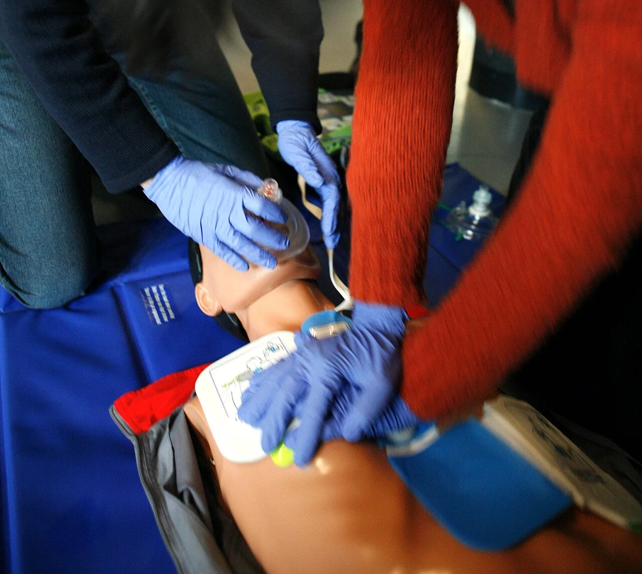
Second, when sudden cardiac arrest occurs, the immediate actions of on-site bystanders determine whether the athlete survives. Every passing minute without chest compressions and defibrillation reduces survival odds by 7 to 10 percent. Schools and community athletic venues must maintain comprehensive Cardiac Emergency Response Plans (CERPs) built upon two essential pillars:
Hands-Only CPR: Upon witnessing an athlete collapse into unresponsiveness with absent or abnormal agonal breathing, bystanders must immediately call emergency services and initiate rhythmic chest compressions. Rhythmic compressions in the center of the chest at 100 to 120 beats per minute to a depth of 2 inches sustain critical coronary perfusion pressure and preserve cerebral cellular integrity.
Automated External Defibrillators (AED): Defibrillation represents the only effective treatment for terminating shockable ventricular fibrillation. Modern AEDs provide voice prompts that guide untrained bystanders through pad placement and rhythm analysis. The device delivers a biphasic electrical shock only when a lethal arrhythmia is identified. When an AED is deployed and discharged within 3 minutes of collapse, athlete survival rates exceed 70 percent [1].

Glossary
Scientific terms used in this article
- Sudden Cardiac Arrest (SCA)
- An abrupt cessation of cardiac pump function triggered by electrical malfunction, leading to immediate circulatory collapse and cerebral hypoperfusion.
- Hypertrophic Cardiomyopathy (HCM)
- A genetic myocardial disorder defined by asymmetric left ventricular hypertrophy, myocyte disarray, and microvascular ischemia predisposing to lethal arrhythmias.
- Ventricular Fibrillation (VF)
- A chaotic, uncoordinated ventricular rhythm exceeding 300 depolarizations per minute, rendering the ventricles incapable of mechanical stroke volume.
- Ion Channelopathy
- An inherited genetic alteration in myocardial ion channel subunits (sodium, potassium, calcium) that destabilizes repolarization without macroscopic structural defects.
- Cardiopulmonary Resuscitation (CPR)
- An emergency lifesaving intervention combining rhythmic chest compressions and rescue ventilations to maintain partial oxygenated coronary and cerebral perfusion.
- Automated External Defibrillator (AED)
- A portable electronic device that autonomously diagnoses shockable ventricular tachyarrhythmias and delivers therapeutic biphasic electrical countershocks.
Sudden Death in the Young: Information for the Primary Care Provider
- Journal
- Pediatrics
- Study Type
- Review
- Year
- 2021
DOI10.1542/peds.2021-052044
Frequently Asked Questions
Common reader questions about this study
What medical screening should student athletes receive before competitive sports?
Every student athlete requires a comprehensive personal and three-generation family medical history alongside a physical examination. If the student reports exertional dizziness, chest tightness, or a family history of premature sudden death before age 50, clinicians must order a 12-lead electrocardiogram (ECG) and echocardiogram.
How can bystanders distinguish sudden cardiac arrest from a seizure when an athlete collapses?
Abrupt cerebral hypoperfusion after cardiac arrest frequently causes brief myoclonic twitching that mimics epileptic seizures. However, the patient will have no carotid pulse and will display absent or abnormal agonal breathing. Bystanders must immediately initiate chest compressions rather than waiting.
Which runners require cardiac screening before registering for a marathon?
Sedentary individuals, runners older than 35, and anyone with existing cardiovascular risk factors (such as hypertension or unmanaged lipid panels) should undergo clinical evaluation and exercise treadmill testing before embarking on distance endurance regimens.
Which symptoms during physical training should prompt immediate medical evaluation?
Exertional syncope (fainting during or immediately following exercise), disproportionate dyspnea, radiating retrosternal chest tightness, and sudden unprovoked palpitations demand immediate training suspension and cardiac workup.
How quickly must CPR and defibrillation occur after an athlete collapses?
Every passing minute without chest compressions and defibrillation drops the odds of survival by approximately 10 percent. Delivering high-quality CPR and an automated external defibrillator shock within 3 to 5 minutes offers survival rates exceeding 70 percent.



No comments yet. Be the first to join the conversation.