
Key Findings
- Over 80 percent of all strokes are completely preventable through clinical blood pressure management, lifestyle modification, and cardiovascular risk reduction.
- Ischemic stroke accounts for approximately 87 percent of cases and is caused by arterial thromboembolism, whereas hemorrhagic stroke accounts for 13 percent and is triggered by vessel rupture.
- Maintaining resting blood pressure under 120/80 mmHg represents the single most powerful clinical intervention to prevent microvascular cerebral damage and aneurysms.
- Recognizing acute warning signs via the BE-FAST acronym ensures patients reach emergency care within the critical 3 to 4.5-hour thrombolytic golden window.
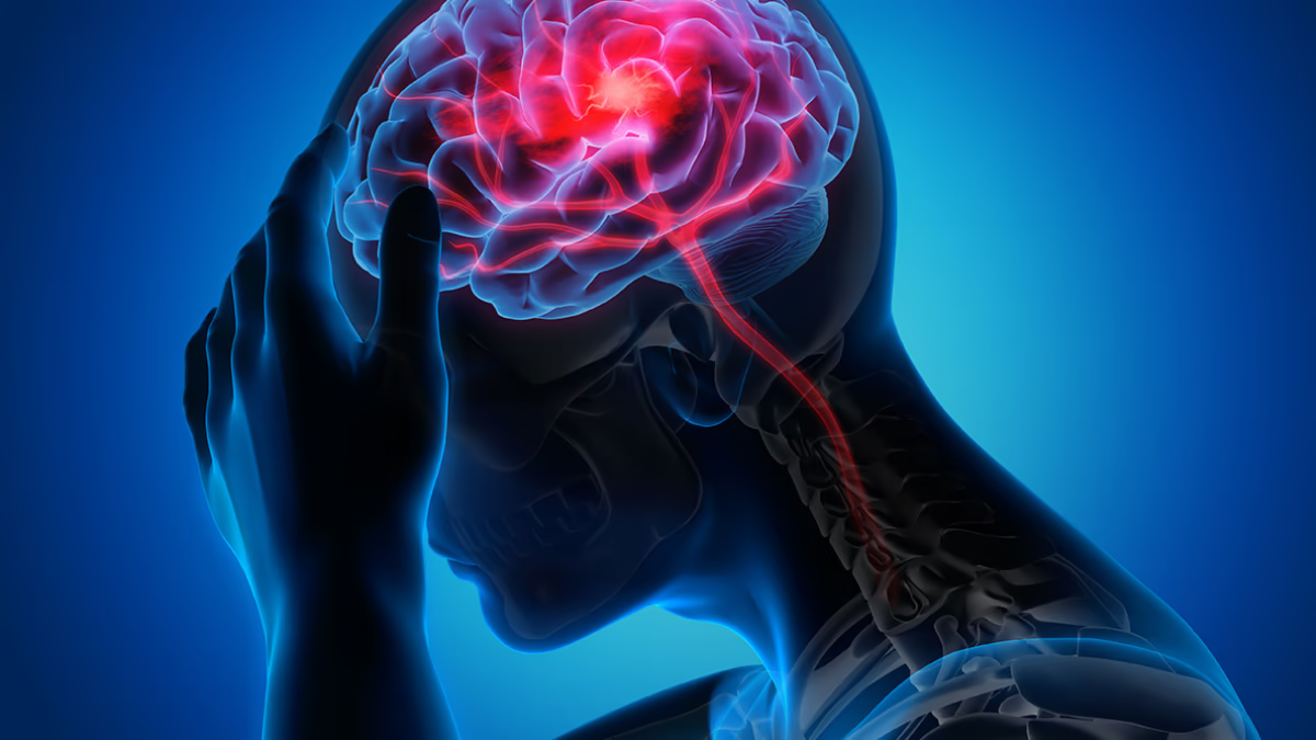
Every forty seconds, someone in the world experiences a stroke, and every three minutes, someone loses their life to this catastrophic cerebrovascular event [1] [5]. As the second leading cause of mortality globally and a primary driver of long-term adult physical disability, a stroke strikes with terrifying suddenness, robbing individuals of speech, mobility, cognition, and independence in a matter of seconds [1] [2]. Yet behind these sobering statistics lies an extraordinary clinical reality: according to the American Heart Association and the World Stroke Organization, over 80 percent of all strokes are completely preventable through targeted medical management and proactive lifestyle modifications [1] [4].
Understanding the physiological mechanics of cerebral circulation, identifying individual risk factors, and implementing evidence-based clinical interventions can safeguard brain health across every stage of adult life [1] [4]. Whether an individual is in their twenties building lifelong vascular resilience or in their seventies managing chronic conditions, science offers clear, actionable pathways to eliminate stroke risk [1] [4].
The Global Stroke Burden and Preventability Reality
The human brain consumes approximately 20 percent of the body’s total oxygen and glucose supply despite accounting for only 2 percent of total body weight [2] [5]. This metabolic demand requires an uninterrupted torrent of oxygenated blood delivered through a delicate network of cerebral arteries [1]. When blood flow is abruptly cut off or when a cranial vessel ruptures, deprived neuronal tissue begins dying at an alarming rate of nearly 1.9 million neurons per minute [1] [3].
The Global Burden of Disease Study published in The Lancet Neurology revealed that the worldwide lifetime risk of stroke from age 25 onward is approximately 25 percent [2]. However, the study emphasized that the vast majority of this burden is mediated through modifiable metabolic, behavioral, and environmental risk factors [2]. By addressing the underlying drivers of endothelial injury, arterial plaque formation, and microvascular strain, premature stroke events can be largely eradicated from modern public health [1] [2].
Ischemic vs Hemorrhagic Stroke: Vascular Pathophysiology
Clinically, cerebrovascular accidents are categorized into two primary pathophysiological mechanisms [1]:
Ischemic Stroke (87% of Cases): Occurs when a blood vessel supplying oxygenated blood to a specific brain region becomes physically occluded by a thrombus (blood clot formed locally within an atheromatous cerebral artery) or an embolus (a blood clot or fatty debris that travels from elsewhere in the body, typically the heart or carotid bifurcation) [1] [5]. Deprived of cellular respiration, the central ischemic core undergoes rapid necrosis, while surrounding tissue, the ischemic penumbra, remains salvageable if blood flow is swiftly restored [1] [3].
Hemorrhagic Stroke (13% of Cases): Occurs when a structurally weakened cerebral blood vessel bursts, spilling pressurized blood into the brain parenchyma (intracerebral hemorrhage) or the subarachnoid space (subarachnoid hemorrhage) [1] [5]. The expanding hematoma compresses adjacent brain tissue, compromises microcirculation, and dramatically elevates intracranial pressure, requiring emergency neurosurgical intervention or aggressive blood pressure reduction [1].
Clinical Comparison: Ischemic vs Hemorrhagic Stroke
| Clinical Metric | Ischemic Stroke (Clot Occlusion) | Hemorrhagic Stroke (Vessel Rupture) | Prevention Priority |
|---|---|---|---|
| Incidence Frequency | Accounts for ~85% to 87% of all stroke events | Accounts for ~13% to 15% of all stroke events | Atherosclerosis and thrombosis prevention addresses the majority |
| Underlying Cause | Atherosclerotic plaque rupture or cardioembolic clot | Chronic hypertension, cerebral aneurysm, or AVM rupture | Managing blood pressure protects against both pathology types |
| Emergency Therapy | IV Thrombolysis (tPA) & mechanical thrombectomy | Surgical hematoma evacuation & intracranial pressure control | Immediate emergency neuroimaging (CT scan) guides differentiation |
| Primary Risk Factor | Elevated LDL-C, diabetes, atrial fibrillation, smoking | Severe unmanaged hypertension, heavy alcohol, coagulopathy | Targeted lifestyle adjustments eliminate shared vascular risks |
| Reversibility Window | Golden 3 to 4.5 hours for IV clot-busting medication | Immediate critical medical care within minutes to hours | Rapid BE-FAST recognition prevents catastrophic permanent deficits |
The Eight Proven Pillars of Primary Stroke Prevention
Extensive clinical trials, including the authoritative American Heart Association and American Stroke Association guidelines, have identified eight foundational pillars that eliminate the root causes of cerebrovascular disease [1] [4]:
1. Optimize Blood Pressure: Maintain resting systolic blood pressure below 120 mmHg and diastolic below 80 mmHg [1]. High pressure mechanically stresses vessel bifurcations, accelerating atherosclerosis and micro-aneurysm formation [1] [5].
2. Lower Atherogenic Cholesterol: Target Low-Density Lipoprotein Cholesterol (LDL-C) below 70 mg/dL for individuals with cardiovascular risk [1]. Using statins or PCSK9 inhibitors stabilizes vulnerable plaques in carotid and intracranial arteries, preventing rupture and embolization [1].
3. Manage Blood Glucose: Maintain Hemoglobin A1c below 6.5 to 7.0 percent [1]. Chronic hyperglycemia induces systemic endothelial dysfunction, thickens capillary basement membranes, and doubles stroke risk [1] [2].
4. Cease Tobacco Use: Smoking doubles ischemic stroke risk and quadruples hemorrhagic stroke risk by promoting platelet aggregation, elevating carbon monoxide, and damaging vascular endothelium [1] [4]. Within two to five years of cessation, an individual’s stroke risk drops to near non-smoker baselines [1].
5. Engage in Regular Physical Activity: Accumulate at least 150 minutes of moderate-intensity aerobic exercise (such as brisk walking, swimming, or cycling) weekly [1] [4]. Exercise promotes vascular nitric oxide release, enhances collateral circulation, and improves metabolic flexibility [1].
6. Adopt a Mediterranean or DASH Dietary Pattern: Emphasize leafy greens, cruciferous vegetables, whole legumes, extra virgin olive oil, nuts, and oily cold-water fish while limiting sodium intake to less than 2,000 mg daily [1] [4]. Dietary potassium and polyphenols actively protect arterial elasticity [1].
7. Maintain a Healthy Body Composition: Reduce visceral abdominal adiposity [1] [2]. Visceral fat secretes pro-inflammatory cytokines that exacerbate arterial stiffening and peripheral insulin resistance [1].
8. Screen for and Treat Atrial Fibrillation: Detect cardiac arrhythmias early through routine pulse palpation or electrocardiograms (ECG) [1]. Anticoagulation therapy reduces cardioembolic stroke risk by over 65 percent in patients with diagnosed atrial fibrillation [1] [5].
Hypertension: The Silent Driver of Cerebrovascular Damage
Among all modifiable risk factors, hypertension is undeniably the single most destructive force in cerebrovascular medicine [1] [2]. Over 50 percent of all ischemic strokes and more than 70 percent of hemorrhagic strokes are directly attributable to uncontrolled high blood pressure [1] [5].
Because high blood pressure causes zero overt symptoms in its early stages, millions of adults live with untreated vascular damage [1]. Under chronic hydraulic shear stress, the smooth muscle layer of cerebral arterioles undergoes hypertrophy and lipohyalinosis, becoming brittle, narrowed, and prone to microscopic thrombotic occlusion (lacunar infarcts) or Charcot-Bouchard aneurysmal ruptures [1] [2]. Clinical trials confirm that every 10 mmHg reduction in systolic blood pressure reduces overall stroke incidence by a staggering 27 to 30 percent [1].
Atrial Fibrillation and Cardioembolic Occlusion
Another major driver of devastating ischemic strokes is Atrial Fibrillation (AFib), the most common cardiac arrhythmia affecting older adults [1] [5].
During atrial fibrillation, the upper cardiac chambers beat chaotically, losing their coordinated contractile squeeze [5]. Blood pools sluggishly within the left atrial appendage, facilitating the rapid assembly of fibrin-rich blood clots [1] [5]. When dislodged, these massive cardioembolic clots pass into the ascending aorta and travel directly into the middle cerebral artery, occluding major blood vessels and causing massive hemispheric strokes [1]. Modern Direct Oral Anticoagulants (DOACs), such as apixaban and rivaroxaban, provide safe, highly effective stroke prophylaxis without the intensive dietary restrictions associated with traditional warfarin [1].
BE-FAST Emergency Recognition and the Golden Treatment Window
When an acute stroke occurs, every passing minute determines whether a patient recovers full neurological function or suffers permanent physical disability [1] [3]. Recognizing acute symptoms using the validated BE-FAST acronym is crucial [1] [4]:
• B – Balance: Sudden loss of balance, dizziness, unsteadiness, or difficulty walking [1].
• E – Eyes: Sudden loss of vision in one or both eyes, or sudden double vision [1].
• F – Face: Facial asymmetry or one side of the face drooping when smiling [1].
• A – Arms: Arm weakness or numbness; one arm drifts downward when raised [1].
• S – Speech: Slurred speech, inability to repeat simple sentences, or difficulty comprehending speech [1].
• T – Time: Time is critical; call emergency medical services immediately to reach an accredited comprehensive stroke center [1] [4].
Patients who arrive at emergency stroke centers within the 3 to 4.5-hour golden window are eligible for intravenous thrombolysis (tissue plasminogen activator / tPA or tenecteplase), which enzymatically dissolves the occluding clot [1]. For large vessel occlusions, endovascular mechanical thrombectomy, where a neuro-interventionalist navigates a micro-catheter to extract the clot physically, can restore perfusion up to 24 hours after onset in selected patients, dramatically reversing neurological deficits [1] [3].
Lifestyle Medicine: Mediterranean Diet, Exercise, and Sleep
Beyond medications, daily lifestyle choices exert continuous epigenetic and physiological control over vascular integrity [1] [4].
Clinical trials like PREDIMED demonstrate that adopting a Mediterranean diet supplemented with extra virgin olive oil or mixed nuts reduces stroke incidence by up to 30 percent compared to low-fat diets [1] [4]. The rich polyphenols, monounsaturated oleic acids, and antioxidant lignans inhibit LDL oxidation, dampen systemic arterial inflammation, and promote endothelial nitric oxide bioavailability [1] [4].
Furthermore, managing obstructive sleep apnea (OSA) is essential [1]. Untreated sleep apnea causes nocturnal hypoxemia and surges of sympathetic catecholamines that spike overnight blood pressure, tripling stroke risk [1]. Prioritizing seven to eight hours of restorative sleep, minimizing chronic psychosocial stress, and maintaining lifelong physical activity form an impenetrable barrier protecting the human brain from vascular catastrophe [1] [4].
Frequently Asked Questions
Primary Sources and References
2021 Guideline for the Prevention of Stroke in Patients With Stroke and Transient Ischemic Attack
- Journal
- Stroke
- Study Type
- Clinical Guideline
- Year
- 2021
DOI10.1161/STR.0000000000000375
Frequently Asked Questions
Common reader questions about this study
What is the single most important factor in preventing a stroke?
Strict blood pressure control is the single most critical preventive measure. Chronic hypertension weakens and stiffens cerebral arteries, dramatically increasing the probability of both ischemic vessel occlusion and hemorrhagic rupture.
What is the difference between an ischemic stroke and a hemorrhagic stroke?
An ischemic stroke occurs when a blood clot (thrombus or embolus) physically obstructs a cerebral artery, depriving downstream brain tissue of oxygen. A hemorrhagic stroke occurs when a weakened cerebral blood vessel bursts, spilling blood into brain parenchyma and creating destructive intracranial pressure.
What does the BE-FAST stroke recognition acronym stand for?
BE-FAST stands for: Balance (sudden loss of balance or coordination), Eyes (sudden vision loss or double vision), Face (facial drooping on one side), Arms (arm weakness or inability to raise both arms), Speech (slurred speech or difficulty finding words), and Time (call emergency medical services immediately).
What is the 'Golden Window' for acute ischemic stroke treatment?
The golden window refers to the first 3 to 4.5 hours following symptom onset. Administering intravenous thrombolytic medications (such as tPA or tenecteplase) within this timeframe dissolves the occluding clot, restoring cerebral blood flow and preventing permanent neurological deficits.
How does atrial fibrillation increase the risk of an ischemic stroke?
Atrial fibrillation causes the heart's upper chambers to quiver irregularly rather than contract forcefully. This sluggish, turbulent blood flow promotes thrombus formation in the left atrial appendage. If a clot breaks loose, it travels directly up the carotid arteries into the brain, causing a massive embolic stroke.
References
- ↩ Kleindorfer, D. O., Towfighi, A., Chaturvedi, S., et al. (2021). 2021 Guideline for the Prevention of Stroke in Patients With Stroke and Transient Ischemic Attack: A Guideline From the American Heart Association/American Stroke Association. Stroke, 52(7), e364-e467.
- ↩ Feigin, V. L., Stark, B. A., Johnson, C. O., et al. (2021). Global, regional, and national burden of stroke and its risk factors, 1990–2019: a systematic analysis for the Global Burden of Disease Study 2019. The Lancet Neurology, 20(10), 795-820.
- ↩ Nogueira, R. G., Jadhav, A. P., Haussen, D. C., et al. (2018). Thrombectomy 6 to 24 Hours after Stroke with a Mismatch between Deficit and Infarct. New England Journal of Medicine, 378(1), 11-21.
- ↩ McLernon, S. (2025). Eight ways to reduce your stroke risk – no matter what age you are. The Conversation, Health Review, 251524.
- ↩ Benjamin, E. J., Muntner, P., Alonso, A., et al. (2019). Heart Disease and Stroke Statistics—2019 Update: A Report From the American Heart Association. Circulation, 139(10), e56-e528.

No comments yet. Be the first to join the conversation.