


Key Findings
- Brain tissue itself has no pain receptors; headache pain originates in meninges, dural vessels, and trigeminal nerves [1].
- Migraine affects over 1 billion people globally (~14% prevalence) and ranks second worldwide in years lived with disability [2].
- Blocking calcitonin gene-related peptide (CGRP) halts trigeminal pain signaling without constricting coronary heart arteries [1, 3].
Brain tissue itself feels zero pain during a splitting headache. Surgeons can touch cerebral cortex tissue in awake patients without causing pain sensation [1]. Instead, headache pain fires from pain-sensitive meninges, dural blood vessels, and cranial nerves wrapped around the brain, chiefly the trigeminal nerve [1, 3].
Migraine affects over one billion people worldwide and roughly 14 percent of the global population [2]. It ranks second globally among all causes of years lived with disability, and first among young women [2].
Decades of bench and clinical neuroscience traced that pain wave to a specific neuropeptide called calcitonin gene-related peptide (CGRP) [1, 3]. Understanding trigeminal signaling and cortical excitability transforms migraines from vague vascular headaches into treatable brain network disorders [1, 2, 3].
Why This Matters: Stopping Disability and Rebound Pain
Most headache sufferers rely solely on over-the-counter painkillers without knowing which neural pathway caused the attack. Treating a complex neurological sensory storm like a simple muscle ache leaves millions under-treated or trapped in recurring attacks [2]. Around the world, missed workdays and impaired concentration during peak adult careers turn untreated headaches into a massive public health burden [2].
Targeted science changed migraine medicine completely over the past decade [3]. Rather than squeezing every blood vessel indiscriminately across the body, modern CGRP receptor antagonists and antibodies interrupt the trigeminal pain messenger selectively [1, 3]. Accurate diagnosis helps patients move from emergency painkiller rescue toward steady preventive care [2, 3].
Daily nervous system habits also shape headache thresholds strongly. Poor sleep hygiene and midnight waking sensitize trigeminal brainstem nuclei, as explored in our guides on how sleep clears brain metabolites and why night awakening alters brain chemistry.
How the Study Was Done: Bench to Clinic Neurobiology
Three major peer-reviewed syntheses map modern headache science from molecular receptors to global healthcare delivery [1, 2, 3]. First, Peter Goadsby and colleagues published an exhaustive sensory neurobiology review in Physiological Reviews in 2017 [1]. They integrated human functional neuroimaging, trigeminal electrophysiology, and brainstem sensory mapping [1].
Second, Lars Edvinsson and colleagues traced over thirty years of CGRP bench-to-clinic translational medicine in Nature Reviews Neurology in 2018 [3]. They followed laboratory measurements of cranial neuropeptide release during migraine attacks through randomized clinical trials of monoclonal antibodies and oral gepants [3].
Third, Messoud Ashina and co-authors synthesized Global Burden of Disease epidemiological data and health-system outcomes in The Lancet in 2021 [2]. Together, these datasets establish both biological mechanisms and population disability metrics [1, 2, 3].
Why Brain Tissue Cannot Hurt: The Trigeminovascular System
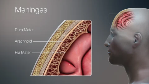
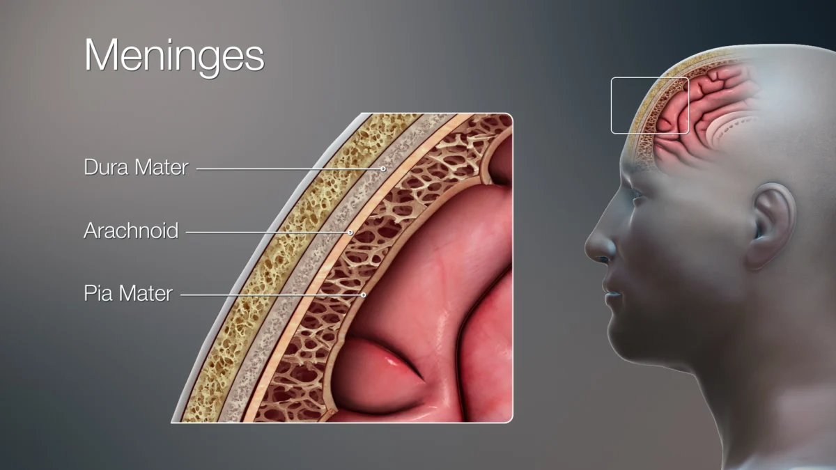
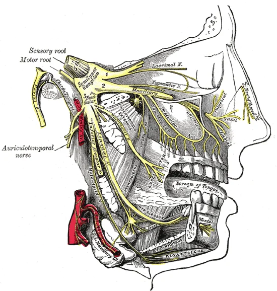
Inside the skull, nerve endings capable of sensing pain sit along three main structures: the tough dura mater membrane, major meningeal arteries, and large venous sinuses [1]. First-order sensory fibers from the ophthalmic division of the trigeminal nerve innervate nearly all of these pain-sensitive coverings [1, 3].
When activated, trigeminal nerve fibers carry pain signals inward to the trigeminal cervical complex inside the brainstem [1]. From there, second-order neurons relay signals upward to the thalamus and sensory cortex, creating throbbing pain behind the eyes, forehead, or temples [1].
At the same time, trigeminal nerve endings release CGRP onto dural blood vessels and nearby immune mast cells [1, 3]. CGRP dilates meningeal arteries and amplifies local neurogenic signaling, locking the pain circuit into a self-reinforcing loop [1, 3].
Why do many people feel throbbing pain worsen every time they climb stairs or bend down? Mechanical stretching of sensitized dural blood vessels fires already-irritable trigeminal nociceptors with every arterial pulse wave [1]. Even subtle fluctuations in blood pressure or cerebrospinal fluid pressure inside the skull send fresh pain volleys along inflamed ophthalmic nerve fibers [1].
How Migraine Unfolds: Aura, Brainstem, and CGRP
Migraine is far more than a dilated blood vessel. Functional imaging captures deep hypothalamus and brainstem changes hours before head pain even starts, explaining premonitory yawning, neck stiffness, and light sensitivity [1].
Roughly one in three migraine patients also experiences visual or sensory aura before pain begins [1, 2]. Neuroscientists link aura to cortical spreading depression, a slow electrophysiological wave sweeping across visual cortex neurons followed by transient neural suppression [1]. As that wave creeps over visual processing regions at millimeters per minute, patients perceive shimmering crescent arcs, blind spots, or tingling pins-and-needles creeping across one hand and face [1].
Premonitory symptoms often arrive hours before aura or head pain starts [1]. Brainstem imaging studies reviewed by Goadsby and colleagues detected early activation in the hypothalamus, periaqueductal gray, and dorsal pons well ahead of headache onset [1]. That deep hypothalamic shift explains why migraine patients report unprovoked yawning, neck tightness, food cravings, or fluid retention long before throbbing pain hits [1].
How a Migraine Attack Amplifies Inside Cranial Nerves
- Phase 1
Hypothalamic Premonitory Shift
Altered brainstem and hypothalamic excitability triggers early fatigue, cravings, and sensory sensitivity [1]. - Phase 2
Cortical Spreading Depression
In aura migraine, a slow wave of neuronal depolarization crosses cortex tissue, generating shimmering visual zigzags [1].
Extended screen glare and disrupted visual ergonomics can aggravate sensory sensitivity during premonitory phases. See our cognitive neuroscience analysis of excessive digital screen exposure and neural pathways alongside our guide on how restorative sleep stabilizes brain excitability.
Tension vs Migraine vs Cluster Headaches
Not all headaches share migraine biology [1, 2]. Tension-type headache feels like a steady, bilateral band around both temples without nausea or severe light aversion [2]. It involves peripheral pericranial muscle tenderness combined with mild central pain facilitation rather than acute trigeminal CGRP surges [1, 2].
Cluster headache lies at the extreme autonomic end of the spectrum [1, 2]. Attacks last 15 minutes to 3 hours, strike strictly behind one eye with tearing or nasal congestion, and follow circadian clock rhythms controlled by the posterior hypothalamus [1]. Patients typically pace restlessly during cluster attacks, unlike migraine sufferers who prefer dark, silent rooms and stillness [1, 2].
Despite causing severe workplace and school disruption, migraine remains widely underdiagnosed in primary care clinics globally [2]. Ashina and colleagues emphasize that straightforward clinical history questions about light sensitivity, nausea, and functional impairment identify true migraine accurately in most patients without specialized scans [2].
Table 1 distinguishes primary headache disorders by pain character, autonomic signs, neurobiology, and clinical response [1, 2, 3].
| Headache Disorder | Typical Pain Pattern | Biological Driver | Distinguishing Features |
|---|---|---|---|
| Migraine [1, 2, 3] | Throbbing, often one-sided | Trigeminal CGRP release and sensory network shift | Light/sound sensitivity, nausea, optional visual aura |
| Tension-Type Headache [1, 2] | Steady, pressing bilateral band | Pericranial muscle tenderness and central facilitation | Mild to moderate intensity; no vomiting or visual aura |
| Cluster Headache [1, 2] | Excruciating pain behind one eye | Hypothalamic activation and trigeminal-autonomic reflex | Eye redness, tearing, nasal stuffiness, extreme restlessness |
| Medication-Overuse Headache [2] | Daily diffuse background ache | Central sensitization from frequent acute pills | Develops after taking acute analgesics 10 to 15+ days monthly |
Compiled from Goadsby et al. (2017), Ashina et al. (2021), and Edvinsson et al. (2018).
Targeted Relief: From Triptans to CGRP Blockers
Understanding CGRP reshaped clinical pharmacology for patients who failed older medicines [3]. Traditional triptans stimulate serotonin 5-HT1B and 5-HT1D receptors, constricting cranial vessels and inhibiting neuropeptide release [1, 3]. Because triptans constrict blood vessels broadly, doctors avoid them in patients with coronary artery disease or stroke history [3].
Anti-CGRP therapies solve that vascular limitation cleanly [3]. Monoclonal antibodies bind circulating CGRP peptide or block its receptor directly for preventive monthly or quarterly protection [3]. Approved preventive antibodies such as erenumab, fremanezumab, galcanezumab, and eptinezumab lower monthly migraine days across episodic and chronic patients in clinical trials [3]. Small-molecule oral antagonists called gepants block CGRP receptors acutely without constricting healthy heart arteries [3].
Lifestyle regularity reinforces pharmacological treatment powerfully [2]. Irregular sleep schedules, skipped meals, sudden caffeine withdrawal, and dehydration act as metabolic stressors on already-excitable hypothalamic networks [1, 2]. Consistent sleep hours, aerobic exercise, and hydration reduce background sensory threshold swings between attacks [2].
Meanwhile, overusing acute pain pills backfires biologically [2]. Taking simple analgesics or combination pain relievers more than 10 to 15 days each month sensitizes central pain pathways, causing daily medication-overuse headaches [2]. Tracking headache frequency and shifting toward targeted prevention stops that rebound cycle [2, 3]. Keeping a simple monthly calendar tracking headache days and pill doses helps doctors distinguish occasional attacks from early medication overuse [2].
What the study cannot tell us: Unanswered Migraine Puzzles
Even with breakthrough CGRP blockers, major scientific questions remain unresolved [1, 3]. Roughly one-third to one-half of chronic migraine patients show incomplete response to CGRP-blocking antibodies alone, proving additional neuropeptides such as PACAP also participate [1, 3].
Neuroscientists also continue investigating why hormonal fluctuations around menstruation amplify trigeminal excitability so strongly in women [2]. Brain imaging also cannot yet provide a single blood test or MRI scan that diagnoses migraine reliably without clinical symptom history [1, 2].
Any sudden, explosive “thunderclap” headache peaking within seconds, or headache accompanied by fever, neck stiffness, weakness, or vision loss, requires emergency medical imaging rather than routine migraine care. Evidence-based self-care and early neurology evaluation prevent years of avoidable disability [2]. Open communication with clinicians about attack frequency, aura symptoms, and family history shortens the path to targeted relief [2, 3].
Frequently Asked Questions About Headaches and Migraine
Frequently Asked Questions
If the brain has no pain receptors, why do headaches hurt so much?
Brain tissue itself lacks nociceptors, yet the surrounding meninges, dural blood vessels, and venous sinuses are richly wired with pain-sensing fibers from the trigeminal nerve. When stimulated, they transmit intense throbbing pain to sensory processing centers.
Is migraine just a severe stress headache?
No. Tension headaches involve mild to moderate bilateral muscle pressure without nausea, whereas migraine is an inherited neurovascular network disorder driven by brainstem excitability, cortical spreading depression, and trigeminal CGRP release.
How do newer CGRP migraine medicines differ from triptans?
Triptans constrict cranial blood vessels through serotonin receptors and cannot safely treat patients with heart disease. CGRP monoclonal antibodies and oral gepants block the CGRP pain pathway directly without systemic vasoconstriction.
Can taking painkillers too often actually cause more headaches?
Yes. Using acute pain relievers or combination tablets on 10 to 15 or more days per month can trigger medication-overuse headache, converting episodic headaches into near-daily pain.
Technical Terms and Definitions
Scientific terms used in this article
- Trigeminovascular SystemCranial Pain Nerve Network
- Trigeminal nerve fibers wrapping pain-sensitive blood vessels inside the outer membranes covering the brain.
- Cortical Spreading DepressionMigraine Aura Wave
- A slow wave of electrical depolarization moving across the cerebral cortex that produces visual or sensory aura.
- MeningesProtective Brain Membranes
- Three tissue layers covering the brain and spinal cord; the outer dura mater contains dense pain fibers.
- GepantsOral CGRP Receptor Blockers
- Small-molecule migraine drugs that block CGRP receptors without constricting coronary blood vessels.
Scientific References and Primary Literature
Pathophysiology of Migraine: A Disorder of Sensory Processing
- Journal
- Physiological Reviews
- Study Type
- Review
- Year
- 2017
DOI10.1152/physrev.00034.2015
Timeline
- 2017
Sensory Processing Model Unified
Goadsby et al. mapped brainstem and hypothalamic premonitory pathways behind migraine.
- 2018
CGRP Therapies Reach Clinic
Edvinsson et al. reviewed clinical translation of anti-CGRP antibodies and oral gepants.
- 2021
Global Disability Measured
Ashina et al. synthesized global data showing over 1 billion people affected worldwide.
References
- ↩ Goadsby, P. J., Holland, P. R., Martins-Oliveira, M., Hoffmann, J., Schankin, C., & Akerman, S. (2017). Pathophysiology of Migraine: A Disorder of Sensory Processing. Physiological Reviews 97(2), 553-622.
- ↩ Ashina, M., Katsarava, Z., Do, T. P., et al. (2021). Migraine: epidemiology and systems of care. The Lancet 397(10283), 1485-1495.
- ↩ Edvinsson, L., Haanes, K. A., Warfvinge, K., & Krause, D. N. (2018). CGRP as the target of new migraine therapies: successful translation from bench to clinic. Nature Reviews Neurology 14(6), 338-350.


No comments yet. Be the first to join the conversation.