



The human eye is an extraordinary optical instrument, yet its evolutionary architecture possesses fundamental physical constraints. Under optimal daylight illumination and high contrast, the resolving power of the human retina reaches an angular limit of approximately one arcminute, translating to a linear spatial resolution of roughly 0.1 millimeters, or 100 micrometers, at the standard reading distance of 25 centimeters. Any biological structure, cellular organelle, bacterial pathogen, or inorganic micro-crystal smaller than this threshold blurs into indistinguishable homogeneity. For millennia, humanity inhabited a reality where the fundamental agents of infectious disease, tissue cellular architecture, and metabolic catalysis remained completely invisible, shrouded behind the impenetrable curtain of optical physics.
The dawn of optical microscopy in the late seventeenth century shattered this perceptual cage. When pioneering Dutch microscopist Antonie van Leeuwenhoek hand-ground tiny, highly spherical biconvex glass beads capable of magnifying specimens up to 275 times, he observed what he famously termed ‘animalcules’ swimming in a single droplet of standing pond water. For the first time, human consciousness bore witness to living single-celled protozoa, bacteria, human erythrocytes, and spermatozoa. Yet, visual observation alone was transient and heavily susceptible to human sketching bias. The true revolution occurred two centuries later with the marriage of optical lenses and photochemical emulsion plates, birthing the discipline of photomicrography [1].
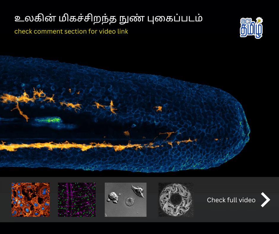
Photomicrography is neither simple photography nor mere laboratory documentation; it represents the precise convergence where advanced optical physics, quantum photonics, fluorescent biochemistry, and artistic composition intersect. Today, modern scientific imaging allows investigators to photograph cellular processes not only at the micrometer scale, but across nanometer spatial dimensions and millisecond temporal scales. Competitions such as the Nikon Small World contest celebrate this spectacular synthesis, revealing that beneath the macro-scale reality we take for granted lies a breathtaking, structurally intricate cosmos vibrating with chromatic brilliance and kinetic vigor.
- 0.1 mm
- Human Eye Limit
- 200 nm
- Abbe Limit
- 10-20 nm
- Super-Resolution
- 0.1 nm
- TEM Resolution
Ernst Abbe and the Classical Diffraction Limit of Light Microscopy
To understand how microscopes capture images of the microscopic realm, one must first confront the wave nature of electromagnetic radiation. In a compound optical microscope, visible light passes through a specimen, enters an objective lens, and is focused to create an enlarged real intermediate image, which is then further magnified by an eyepiece or recorded directly onto a digital complementary metal-oxide-semiconductor (CMOS) sensor. In 1873, German physicist Ernst Abbe established the fundamental mathematical relationship governing optical resolution, forever changing microscopy.
Abbe discovered that light passing through a tiny specimen aperture does not travel in infinitely sharp straight rays; rather, it undergoes wave diffraction, bending and forming an interference pattern consisting of a central bright disk surrounded by concentric dark and bright rings. This optical phenomenon is known as the Airy disk. When two adjacent points in a microscopic sample are positioned closely together, their corresponding Airy diffraction disks overlap. If the centers of the two disks are closer than the radius of the central bright peak, the two distinct points merge into a single unresolved blur.
Mathematically, Abbe formalized the minimum resolvable distance (d) between two distinguishable points as: d = lambda / (2 * NA) Here, lambda represents the wavelength of the illuminating light, and NA denotes the numerical aperture of the objective lens, defined as NA = n * sin(alpha), where n is the refractive index of the immersion medium (such as air, water, or specialized cedar immersion oil) and alpha is the half-angle of the maximum cone of light captured by the lens. Because visible light spans wavelengths from approximately 400 nanometers (violet) to 700 nanometers (deep red), and the maximum practical numerical aperture of high-end oil-immersion lenses peaks around 1.4 to 1.45, Abbe’s diffraction limit establishes that conventional optical microscopes cannot discern structural details smaller than approximately 200 nanometers laterally (XY) and 500 nanometers axially (Z).
For over a century, this 200-nanometer diffraction barrier was regarded as an immutable law of nature, akin to the speed of light in vacuum. It meant that while researchers could clearly view eukaryotic cells (typically 10 to 30 micrometers in diameter) and large bacteria (1 to 2 micrometers), individual viral particles (20 to 150 nanometers), ribosome protein synthesis factories (25 nanometers), and individual actin filament cables (7 nanometers) remained frustratingly obscured beyond optical resolution.
Fluorescence Microscopy and Confocal Laser Scanning Technology
Standard brightfield transmission microscopy suffers from a profound limitation when imaging biological tissues: most living cells are composed primarily of water, electrolytes, and transparent organic polymers, rendering them virtually colorless and devoid of optical contrast. While chemical dyes such as hematoxylin and eosin can stain fixed, dead tissues, observing dynamic intracellular physiology demanded an entirely different paradigm. That paradigm emerged with fluorescence microscopy and the discovery of naturally occurring fluorescent proteins.
Fluorescence occurs when an orbital electron within a specialized molecular chromophore (a fluorophore) absorbs a photon of a specific high-energy, shorter excitation wavelength, elevating the electron to an excited singlet quantum state. As the molecule relaxes back to its electronic ground state through vibrational non-radiative decay, it emits a secondary photon of lower energy and longer wavelength. This energetic wavelength shift between absorbed excitation light and emitted fluorescence is known as the Stokes shift. By employing specialized optical dichroic beam splitters and barrier emission filters, microscopists can completely filter out the intense excitation illumination, allowing only the pure, glowing emitted signal from targeted molecular structures to reach the camera detector.
The true golden age of biological photomicrography arrived with the molecular cloning of Green Fluorescent Protein (GFP), originally isolated from the crystal jellyfish Aequorea victoria. By genetically fusing GFP or its engineered chromatic variants (cyan, yellow, red, and far-red proteins) to specific endogenous gene sequences, molecular biologists can cause living cellular machines to glow with vibrant luminescence without killing the host organism. Similar to natural bioluminescent phenomena explored in the science of cold light across bioluminescent organisms, these fluorescent molecular probes provide astonishing contrast against pitch-black backgrounds.
However, standard widefield epifluorescence microscopy suffers from significant out-of-focus flare. When a specimen has considerable thickness (such as a developing zebrafish embryo or a neural tissue slice), illuminating the entire sample excites fluorescent molecules above and below the focal plane. This out-of-focus emission washes out fine details into a hazy fog. To overcome this hurdle, Marvin Minsky patented the concept of Confocal Laser Scanning Microscopy (CLSM) [2].
CLSM achieves optical sectioning by placing a physical pinhole aperture conjugate to the focal plane directly in front of the photon detector. Laser beams of precise wavelengths scan across the specimen point by point. While light originating precisely from the in-focus focal plane passes cleanly through the conjugate pinhole onto a high-sensitivity photomultiplier tube (PMT) or avalanche photodiode, out-of-focus rays originating from deeper or shallower depths strike the edges of the pinhole mask and are physically blocked. By translating the microscope objective axially in micro-stepping increments of 0.1 micrometers, automated confocal systems acquire an array of razor-sharp optical sections. Sophisticated volumetric algorithms then assemble these two-dimensional optical slices into breathtaking, interactive three-dimensional digital reconstructions of intact living tissues.
Breaking the Diffraction Limit: Super-Resolution Nanoscopy
By the late twentieth century, scientists recognized that while Abbe’s diffraction limit held true for classical linear optics, quantum photophysics offered ingenious mechanisms to circumvent the wave barrier. This realization gave birth to super-resolution nanoscopy, an achievement recognized with the 2014 Nobel Prize in Chemistry awarded to Stefan Hell, Eric Betzig, and W.E. Moerner.
Stefan Hell pioneered Stimulated Emission Depletion (STED) microscopy [3]. In a STED system, a focused circular laser beam excites fluorophores within a standard diffraction-limited focal spot (approximately 250 nanometers wide). Simultaneously, a second, red-shifted laser beam passed through a phase plate forms a donut-shaped ring around the central excitation spot. This donut beam operates through stimulated emission: any excited fluorophore struck by this depleting laser is instantly forced back to its ground state before it can emit spontaneous fluorescence. Consequently, fluorescence is physically silenced everywhere except within the microscopic sub-diffraction hollow center of the donut beam, compressing the effective point spread function down to an astonishing 20 to 30 nanometers.
Simultaneously, Eric Betzig and Samuel Hess developed single-molecule localization microscopy techniques, widely known as Photoactivated Localization Microscopy (PALM) and Stochastic Optical Reconstruction Microscopy (STORM) [2]. These methodologies rely on photo-switchable fluorescent molecules that can be selectively toggled on and off with low-intensity ultraviolet light. Rather than exciting all fluorophores simultaneously into an overlapping optical blur, PALM activates only a sparse, randomly distributed subset of isolated molecules in each camera frame.
Because each glowing molecule in a frame is separated from its neighbors by several micrometers, its individual diffraction pattern can be fitted to a mathematical two-dimensional Gaussian distribution function. By calculating the exact centroid coordinate of thousands of individual photon bursts across tens of thousands of consecutive video frames, a computational algorithm renders an ultra-dense coordinate map of molecular positions. Super-resolution microscopy effectively pushed optical resolution down to 10 to 20 nanometers, allowing direct visualization of individual nuclear pore complexes, viral capsid assemblies, and neurotransmitter receptor clustering at synaptic junctions.
Electron Microscopy: Harnessing de Broglie Matter Waves
While advanced super-resolution optical nanoscopy provides unmatched molecular specificity within fluorescently labeled cells, it cannot image the dense, unstained context of overall ultrastructure. To visualize specimens at sub-nanometer and atomic scales, physicists stepped beyond electromagnetic photons entirely, turning to accelerated electron beams and quantum matter waves.
In 1924, French physicist Louis de Broglie formulated the revolutionary hypothesis of wave-particle duality, proposing that all material particles possess an associated wavelength inversely proportional to their momentum: lambda = h / p where h is Planck’s constant and p is particle momentum. When electrons are accelerated across high-voltage electrical potentials (ranging from 80 kilovolts to 300 kilovolts) within an ultra-high vacuum column, their relativistic de Broglie wavelength drops below 0.005 nanometers (0.05 Ångströms), over 100,000 times shorter than visible light photons. Guided and focused not by glass lenses, but by electromagnetic coils and magnetic apertures, electron beams unlocked the field of electron microscopy.
Electron microscopy branches into two primary analytical architectures: Transmission Electron Microscopy (TEM) and Scanning Electron Microscopy (SEM). In TEM, an accelerated electron beam transmits directly through an ultrathin section of plastic-embedded tissue (typically 50 to 70 nanometers thick) stained with heavy metal salts such as uranyl acetate and lead citrate. As electrons scatter against electron-dense heavy atoms, they create high-contrast projection images on a scintillating detector, resolving lipid bilayer membranes, mitochondrial cristae, and ribosomal subunits with atomic precision.
Conversely, SEM provides spectacular three-dimensional surface topographical imaging. In SEM, a finely focused electron probe rasters across the surface of a specimen coated with an ultra-thin conductive layer of gold-palladium or platinum. The primary electron beam knocks out low-energy secondary electrons from the surface topography of the sample. An Everhart-Thornley detector collects these secondary electrons, translating the signal intensity into dramatic, high-depth-of-field images of insect compound eyes, pollen grains, microchips, and fungal spores. Cryogenic electron microscopy (Cryo-EM) has pushed this frontier even further, rapidly vitrifying biological samples in liquid ethane at minus 180 degrees Celsius to resolve macromolecular protein structures at atomic resolution without crystallization artifacts.
Comparative Architecture: Optical, Confocal, and Electron Microscopy
Selecting the appropriate photomicrographic platform requires balancing resolution, sample preparation constraints, specimen viability, and imaging speed. While optical brightfield remains accessible for education and rapid clinical screening, fluorescence confocal and electron microscopy represent specialized research powerhouses designed for distinct biological and materials inquiries.
| Modality | Illumination Source | Practical Resolution | Specimen Condition | Primary Research Use |
|---|---|---|---|---|
| Brightfield Optical | Broadband Visible Light | ~200 nm (lateral) | Living or fixed cells, stained histology | Routine pathology, tissue morphology, hemocytometry |
| Confocal Laser (CLSM) | Coherent Solid-State Lasers | ~180 nm lateral, ~500 nm axial | Living cells, fixed tissues with fluorophores | 3D optical sectioning, protein colocalization, live imaging |
| Super-Resolution (STED/PALM) | Pulsed lasers / depletion donuts | 10 to 30 nm | Fluorescently tagged proteins, fixed cells | Synaptic vesicles, cytoskeleton filaments, nuclear pores |
| Scanning Electron (SEM) | Accelerated Electron Beam (1-30 kV) | 1 to 5 nm | Dehydrated, sputter-coated, high vacuum | Surface topography, fractography, nanomaterials |
| Transmission Electron (TEM) | High-Energy Electron Beam (80-300 kV) | 0.1 to 0.2 nm (atomic scale) | Ultrathin sections, cryo-vitrified samples | Viral structures, macromolecular complexes, crystal lattices |
Computational Photomicrography: ImageJ, Deconvolution, and Neural Vision
In modern science, capturing raw photons or electrons onto a detector is merely the first half of the imaging pipeline. The raw digital image produced by a microscope is a mathematical convolution of the specimen’s actual ground-truth structure and the optical system’s Point Spread Function (PSF), the three-dimensional diffraction response of the microscope optics to an infinitely small point source of light. To recover spatial fidelity, computational photomicrography relies heavily on mathematical deconvolution algorithms.
Deconvolution utilizes iterative mathematical calculations, such as the Richardson-Lucy algorithm, to computationally reassign out-of-focus blur photons back to their genuine focal origin. By modeling the known optical aberrations of the objective lens and refractive index mismatches, deconvolution sharpens three-dimensional confocal and widefield image stacks with dramatic precision. The open-source scientific image processing software ImageJ, alongside its modern distribution Fiji, developed by the US National Institutes of Health (NIH), serves as the global standard analytical workbench for executing these complex transforms, quantifying fluorescence intensities, and tracking particle kinetics [1].
Furthermore, artificial intelligence and deep learning convolutional neural networks (CNNs) have revolutionized microscopy workflows. Advanced neural networks trained on paired low-resolution and high-resolution ground truth datasets can perform denoising, blind deconvolution, and content-aware image restoration without damaging fragile biological specimens through excessive laser phototoxicity. Automated algorithms can segment thousands of individual cell nuclei, trace convoluted neuronal axons across gigapixel brain datasets, and classify cellular phenotypes with speeds unachievable by human observers. For a broader contextual framework on scientific categorization and terminology, explore our detailed scientific terminology guide, which maps cross-disciplinary nomenclatures across computational and natural sciences. Similarly, modern machine learning models and automated reasoning pipelines, such as those discussed in our analysis of reasoning architectures in artificial intelligence, are now being deployed directly on microscope workstations for real-time sample navigation and adaptive acquisition.
Biomedical Breakthroughs and Industrial Frontiers Enabled by Microscopy
The transformative impact of modern photomicrography reverberates across virtually every domain of medicine, engineering, and environmental stewardship. In cancer biology, intravital confocal and multiphoton microscopy allow researchers to peer directly into living mammalian tumor microenvironments in real time. Scientists can observe cancer cells detaching from primary solid tumors, squeezing through tight endothelial junctions into capillary vessels (intravasation), and colonizing distant organs in the deadly cascade known as metastasis. Understanding these biomechanical and molecular events has directly accelerated the targeted development of anti-angiogenic and immunotherapy pharmacological agents.
In neurobiology, high-throughput serial section electron microscopy and expansion microscopy are driving the monumental endeavor of connectomics, mapping every synaptic connection within complex brain tissue. By reconstructing thousands of terabytes of electron micrographs, neuroscientists are decoding the physical wiring diagrams that underpin memory storage, sensory perception, and cognitive computation. Meanwhile, developmental biologists utilize light-sheet fluorescence microscopy (LSFM) to track the spatial positioning and differentiation of every single cell in a developing fruit fly or zebrafish embryo over days of continuous non-invasive video recording [4].
Beyond clinical medicine, industrial materials science and semiconductor manufacturing depend fundamentally on photomicrography and electron microscopy. Advanced fabrication facilities (fabs) producing 2-nanometer and 3-nanometer microchip architectures employ high-resolution SEM and atomic force microscopy (AFM) to inspect extreme ultraviolet (EUV) lithography silicon wafers, identifying atomic-scale defect dislocations that could render an entire batch of microprocessors nonfunctional. Similarly, environmental researchers analyze microplastic contamination in ocean water and air samples through polarized light microscopy and micro-Raman spectroscopy, assessing ecological toxicological threats with microscopic precision.
Scientific References and Primary Literature
NIH Image to ImageJ: 25 years of image analysis
- Journal
- Nature Methods
- Study Type
- peer-reviewed
- Year
- 2012
DOI10.1038/nmeth.2089
Frequently Asked Questions
Common reader questions about this study
Can living biological cells survive high-resolution electron microscopy?
No, standard electron microscopy requires specimens to be completely dehydrated, coated with heavy metals, and placed inside an ultra-high vacuum chamber, which destroys living biological processes. To observe living cells in real time, researchers utilize advanced optical methods such as live-cell confocal microscopy, light-sheet microscopy, or phase-contrast imaging.
What is the difference between magnification and resolution in microscopy?
Magnification refers to how much larger an image appears compared to the physical object, whereas resolution is the minimum distance between two distinct points at which they can still be distinguished as separate entities. Empty magnification occurs when an image is enlarged without improving resolution, resulting in a large but blurry image devoid of additional structural detail.
How do fluorescent dyes produce vivid colors in photomicrographs?
Fluorescent dyes do not reflect natural ambient colors. Instead, they absorb short-wavelength excitation light (such as ultraviolet or blue laser lines) and emit longer-wavelength light (such as green, yellow, or deep red) through fluorescence. In multichannel imaging, distinct laser wavelengths excite different fluorophores tagged to specific cellular structures, producing composite chromatic images.
What is numerical aperture and why is it crucial for image quality?
Numerical aperture (NA) measures the range of angles over which an objective lens can accept light rays from a specimen, combined with the refractive index of the immersion medium. A higher numerical aperture captures wider diffraction angles, directly yielding superior resolving power, higher optical sharpness, and significantly brighter images.
Glossary
Scientific terms used in this article
- Photomicrography
- The specialized science and technique of capturing photographs of microscopic subjects through an optical or electron microscope.
- Diffraction Barrier
- The fundamental physical limit established by Ernst Abbe where optical resolution is restricted by the wave nature and wavelength of illuminating light.
- Confocal Microscopy
- An optical imaging technique that employs spatial pinhole filtering to eliminate out-of-focus light in specimens thicker than the focal plane.
- STED Nanoscopy
- Stimulated Emission Depletion microscopy, a super-resolution technique that silences peripheral fluorescence with a donut-shaped depletion laser.
- Deconvolution
- A computational algorithm that reassigns blurred and diffracted out-of-focus photons back to their original point of emission in 3D image stacks.
- Numerical Aperture
- A dimensionless number characterizing the range of angles over which an optical system can accept or emit light.
References
- ↩ Schneider, C. A. et al. (2012). NIH Image to ImageJ: 25 years of image analysis. Nature Methods, 9(7), 671–675.
- ↩ Betzig, E. et al. (2006). Imaging intracellular fluorescent proteins at nanometer resolution. Science, 313(5793), 1642–1645.
- ↩ Hell, S. W., & Wichmann, J. (1994). Breaking the diffraction resolution limit by stimulated emission: stimulated-emission-depletion fluorescence microscopy. Optics Letters, 19(11), 780–782.
- ↩ Stephens, D. J., & Allan, V. J. (2003). Light microscopy techniques for live cell imaging. Science, 300(5616), 82–86.

No comments yet. Be the first to join the conversation.