




Key Findings
- Metabolic dysfunction-associated steatotic liver disease affects up to 11.3 percent of normal-weight individuals across Asia.
- Biopsy records confirm advanced F3-F4 fibrosis in 29.3 percent of lean liver patients, demonstrating severe structural damage despite normal body weight.
- Transient elastography (FibroScan) predicts advanced liver fibrosis with 87 percent diagnostic accuracy, eliminating the need for invasive needle biopsies.
For decades, conventional medical wisdom maintained that fatty liver disease belonged almost exclusively to individuals with visible abdominal weight or high body mass index. Most people assume that staying thin provides an automatic metabolic shield against internal organ damage. Yet routine medical checkups and abdominal ultrasound screenings frequently reveal an unsettling diagnosis: grade 1 or grade 2 fatty liver in adults who run daily, eat moderately, and maintain lean physiques [1].
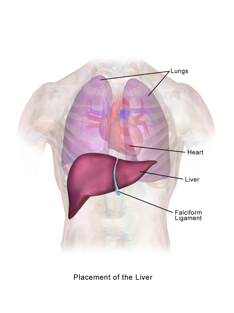
The human liver operates as a central biochemical refinery, processing nutrients, filtering toxins, and regulating systemic energy storage. When fat accumulates inside hepatocytes, body weight tells only a fraction of the story. Known clinically as metabolic dysfunction-associated steatotic liver disease (MASLD), this condition increasingly develops in non-obese individuals. On November 17, 2026, a landmark multinational study published in the Journal of the American Medical Association (JAMA) analyzed 18,326 biopsy-confirmed patients across 41 countries, confirming that lean patients face rates of liver failure, cancer, and mortality nearly identical to overweight individuals once fibrosis sets in [1].
- 18,326
- International Patients Analyzed Comprehensive clinical records from 41 countries
- 29.3%
- Advanced Fibrosis in Lean Cohort Biopsy-confirmed F3-F4 stage scarring
- 11.3%
- Lean MASLD Prevalence in Asia Highest regional rate among normal-weight adults
- 0.87
- FibroScan Accuracy (AUC) Validated non-invasive acoustic wave assessment
The Biology of Hidden Organ Fat in Lean Bodies
Body Mass Index measures external mass relative to height. However, it cannot differentiate between skeletal muscle, subcutaneous fat, and visceral fat. Adipose tissue distributes into two distinct anatomical compartments: subcutaneous fat stored beneath the skin, and visceral fat packed tightly around internal abdominal organs. While subcutaneous fat produces visible body contours, visceral fat acts as an inflammatory metabolic disruptor [1].
Every individual carries a genetically determined personal fat threshold. This evolutionary concept emerged from the adipose tissue expandability hypothesis. Under healthy conditions, subcutaneous fat acts as an expansive buffer. It safely absorbs excess circulating lipids during periods of surplus calorie intake. When subcutaneous fat depots reach their maximal biological storage capacity, adipose stem cells fail to recruit new fat cells. Instead, existing fat cells undergo hypertrophy, recruit inflammatory macrophages, and experience cellular oxygen deprivation [1, 2].
Unable to store under the skin, surplus lipids spill directly into systemic circulation. They settle into vital non-adipose organs such as the liver, pancreas, and deep skeletal muscle bands. Pathologists describe this damaging process as ectopic lipid deposition. In individuals with genetically restricted subcutaneous expandability, even small carbohydrate surpluses trigger rapid lipid overflow into hepatic tissue. Similar hidden systemic lipid anomalies explain why heart attacks strike people with normal cholesterol despite standard blood lipid panels appearing normal [1].
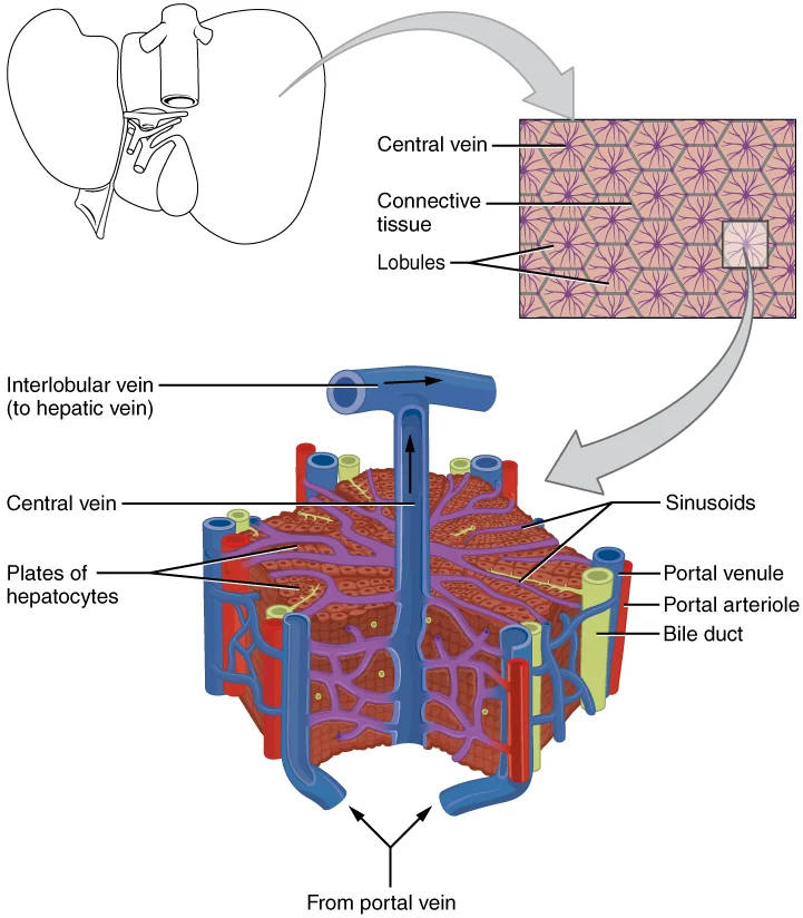
Visceral adipocytes surrounding the mesenteric vascular bed exhibit high rates of lipolysis. They release free fatty acids directly into the portal vein. This anatomical arrangement delivers an unbuffered flood of toxic lipids straight into the liver. Hepatocytes must process these excess fatty acids through mitochondrial beta-oxidation. When beta-oxidation capacity is overwhelmed, hepatocytes store excess fatty acids as neutral lipid droplets, establishing hepatic steatosis [1].
Genetic Architecture and Metabolic Drivers
The development of hepatic steatosis in lean individuals stems from a convergence of genetic susceptibility, altered cellular lipid export, and muscle insulin resistance. Genome-wide association studies identified a major single nucleotide polymorphism in patatin-like phospholipase domain-containing protein 3, known as PNPLA3 rs738409 (I148M). Under normal circumstances, the PNPLA3 enzyme assists in hydrolyzing intrahepatic triglycerides on the surfaces of lipid droplets [1].
The rs738409 missense mutation inhibits proteasomal degradation of the enzyme. This defect creates an inactive protein trap on hepatocyte lipid droplets that halts triglyceride mobilization. In the international JAMA cohort, 48.7 percent of lean MASLD patients carried this variant. This frequency significantly exceeded the 36.2 percent observed in overweight patients [1]. This genetic profile explains why individuals with minimal visible body fat can develop substantial hepatic lipid accumulation.
Additional genetic variants also contribute to lean steatosis. Mutations in TM6SF2 (transmembrane 6 superfamily member 2) impair the secretion of very low-density lipoproteins (VLDL). While this mutation protects cardiovascular arteries by lowering circulating blood lipids, it traps triglycerides inside hepatic parenchyma. Similarly, variations in MBOAT7 disrupt phosphatidylinositol remodeling in cell membranes, increasing susceptibility to liver inflammation [1, 2].
Compounding this genetic predisposition is skeletal muscle insulin resistance driven by physical inactivity. Even without elevated fasting blood glucose, impaired glucose transporter translocation in sedentary muscle fibers diverts dietary carbohydrates toward the liver. Through hepatic de novo lipogenesis, hepatocytes convert this glucose surge into palmitic acid and triglycerides, overwhelming export mechanisms. Clinical observations utilizing continuous glucose monitoring sensors reveal that lean individuals with early insulin resistance often experience large post-meal glucose excursions that go undetected on standard fasting tests [1, 2].
How the Study Was Conducted: Biopsy Cohort Analysis
To evaluate whether lean steatotic liver disease behaves differently from typical obesity-linked liver disease, the Global MASLD Consortium conducted a rigorous retrospective-prospective international cohort investigation. Researchers gathered histopathological and long-term longitudinal clinical records from 18,326 patients across 41 tertiary medical centers in North America, Europe, Asia, Latin America, and the Middle East [1].
Every included participant underwent a definitive percutaneous liver biopsy. Biopsy cores were analyzed by expert hepatopathologists using standardized scoring systems from the Pathology Committee of the NASH Clinical Research Network. Steatosis grade (0 to 3), lobular inflammation (0 to 3), hepatocellular ballooning (0 to 2), and fibrosis stage (F0 to F4) were meticulously recorded [1].
Patients were categorized by World Health Organization BMI classifications. Researchers used the internationally established Asian cut-off of BMI less than 23 kg/m² and non-Asian cut-off of BMI less than 25 kg/m² to identify true lean MASLD. Patients with viral hepatitis (B or C), excessive alcohol consumption, autoimmune liver disorders, or drug-induced steatosis were excluded to ensure rigorous metabolic isolation [1].
Longitudinal clinical outcomes tracked over a median follow-up period of 7.4 years included liver-related clinical events (decompensated cirrhosis, ascites, hepatic encephalopathy, variceal bleeding), development of hepatocellular carcinoma, and all-cause mortality. Statistical analyses applied multivariable Cox proportional hazards models adjusted for age, sex, diabetes status, baseline fibrosis stage, and lipid profiles [1].
Clinical Comparison: Lean Versus Overweight Liver Patients
The comparative findings from the JAMA investigation challenge traditional diagnostic assumptions. While overall disease prevalence is higher in overweight populations, lean individuals diagnosed with MASLD display severe histopathological lesions at remarkably similar rates once the condition initiates [1].
| Clinical Parameter | Lean MASLD (BMI <25) | Non-Lean MASLD (BMI ≥25) | Statistical Significance |
|---|---|---|---|
| Prevalence in Asia | 11.3% | 28.5% | p < 0.001 |
| Advanced Fibrosis (F3-F4) | 29.3% | 34.8% | p = 0.08 (Comparable) |
| Hepatic Steatosis Grade ≥2 | 68.4% | 82.1% | p < 0.01 |
| PNPLA3 rs738409 (G Allele) | 48.7% | 36.2% | p < 0.001 (Higher in Lean) |
| Type 2 Diabetes Comorbidity | 42.1% | 61.8% | p < 0.001 |
| 10-Year Liver Mortality Rate | 4.8% | 5.2% | p = 0.42 (Identical Risk) |
Source: de Avila L et al., JAMA, November 17, 2026. DOI: 10.1001/jama.2026.18491
Crucially, 29.3 percent of lean MASLD patients presented with advanced F3 bridging fibrosis or F4 cirrhosis at their initial diagnostic biopsy. Furthermore, longitudinal survival modeling demonstrated that ten-year liver-related mortality rates between lean patients (4.8%) and non-lean patients (5.2%) showed no statistically significant difference (hazard ratio: 0.94, p = 0.42) once fibrosis severity was matched [1].
Lean patients demonstrated lower absolute rates of arterial hypertension and dyslipidemia than overweight counterparts. However, their rates of cardiovascular events remained elevated compared to healthy controls. This observation underlines that ectopic organ fat generates widespread systemic vascular inflammation, regardless of external waistline measurements [1, 2].
The Three Pathological Stages to Liver Cirrhosis
Steatotic liver disease progresses along a structured pathological continuum. It transitions from simple benign lipid storage to irreversible tissue architecture destruction [1, 2].
Pathological Cascade from Steatosis to Cirrhosis
Stage 1: Hepatic Steatosis
Stage 2: Steatohepatitis (MASH)
Stage 3: Stellate Activation & Fibrosis
Stage 4: Decompensated Cirrhosis
During the steatohepatitis transition, excessive intrahepatic free fatty acids undergo incomplete beta-oxidation inside mitochondria, generating reactive oxygen species. These toxic molecules cause lipid peroxidation of cellular membranes. Ballooning hepatocytes release danger-associated molecular patterns (DAMPs) and inflammatory cytokines like TNF-alpha and interleukin-6 [2, 3].
These cytokines directly activate quiescent hepatic stellate cells located in the space of Disse. Upon activation, stellate cells transdifferentiate into proliferative myofibroblasts. They produce massive quantities of fibrillar collagen types I and III. As collagen bands expand, they bridge portal tracts and central veins, distorting vascular architecture and compressing sinusoidal blood flow [1, 2].
When bridging fibrosis becomes diffuse throughout the parenchyma, regenerative nodules form. This defines cirrhosis. At this advanced juncture, normal hepatic metabolic clearance fails, bile secretion declines, and elevated vascular resistance causes portal hypertension, resulting in ascites and gastroesophageal varices [1].
Study Limitations and Unanswered Questions
While the global JAMA dataset provides unprecedented statistical power, several methodological limitations warrant consideration. First, tertiary academic medical centers contributed all patient cohorts. This setting creates potential referral bias. Lean patients referred for invasive liver biopsy may have possessed higher pre-test likelihoods of advanced disease compared to community-dwelling lean populations [1].
Second, comprehensive dietary macronutrient breakdowns (such as specific ratios of ultra-processed carbohydrates versus saturated fats) and precise objective physical activity tracking through accelerometry were unavailable for retrospective historical subsets. Third, while genetic PNPLA3 screening was completed in major sub-cohorts, emerging regulatory genetic variants such as TM6SF2 and MBOAT7 were not universally sequenced across all participating centers [1].
Fourth, serial follow-up biopsies were not mandated across the entire cohort. Because repeated needle biopsies carry procedural risks, histological resolution or fibrosis progression was monitored through non-invasive surrogates and clinical liver decompensation endpoints rather than routine repeat tissue sampling [1].
Non-Invasive Diagnostic Advances: Beyond Needle Biopsy
For decades, percutaneous needle biopsy remained the sole reliable method to differentiate simple steatosis from progressive fibrosis. However, invasive biopsies carry hemorrhage risks, post-procedural pain, and substantial sampling variability. A standard biopsy needle captures only 1/50,000th of the entire organ volume, creating room for sampling errors [1].

Clinical practice has transitioned toward non-invasive biomarkers. The Fibrosis-4 index combines patient age, platelet count, aspartate aminotransferase, and alanine aminotransferase into an accessible clinical scoring formula. A FIB-4 score below 1.30 provides a 90 percent negative predictive value, safely ruling out advanced fibrosis in primary care settings [1].
When FIB-4 yields indeterminate values, transient elastography (FibroScan) measures low-frequency shear wave velocity through liver parenchyma. Tissue stiffness measured in kilopascals directly correlates with histological fibrosis stage. In clinical validation, FibroScan achieves an area under the ROC curve of 0.87 for detecting advanced F3-F4 fibrosis without surgical intervention [1].
Magnetic Resonance Elastography (MRE) represents an even more sensitive non-invasive modality. By evaluating acoustic shear waves across the entire liver cross-section, MRE accurately stages fibrosis across all grades (F1 through F4). MRE proves particularly valuable in lean patients where acoustic ultrasound penetration might be affected by narrow rib intercostal spacing [1, 2].
Why This Research Matters: Clinical and Public Health Impact
These findings reshape clinical screening guidelines across international hepatology societies. Traditional screening protocols rarely recommend liver evaluation for normal-weight adults unless routine liver enzyme panels display pronounced abnormalities. However, clinical studies show that up to 60 percent of lean individuals with biopsy-proven steatohepatitis maintain normal ALT and AST readings [1].
Public health frameworks must recognize that normal body mass index does not guarantee metabolic wellness. Routine incorporation of non-invasive fibrosis scoring and targeted elastography for patients with visceral adiposity, family history of early diabetes, or persistent fatigue can prevent progression to irreversible cirrhosis decades before clinical liver failure occurs [1, 2].
Furthermore, global drug development trials for steatohepatitis must deliberately recruit lean patients. Currently, many phase 3 clinical trials mandate elevated BMI as an inclusion criterion. This practice excludes lean individuals from accessing emerging therapeutics like thyroid hormone receptor agonists and GLP-1 receptor modulators [1, 2].
Evidence-Based Prevention and Nutritional Reversal
Reversing intrahepatic fat accumulation requires addressing underlying insulin sensitivity and restoring balanced lipid export. Clinical trials show that even a 3 to 5 percent reduction in total body weight achieves substantial reduction in intrahepatic steatosis, while a 7 to 10 percent reduction promotes regression of histologically confirmed fibrosis [2, 3].
Nutritional intervention must prioritize eliminating refined carbohydrates, fructose syrups, and deep-fried processed snacks that drive hepatic de novo lipogenesis. Deep-fried snack foods prepared with repeatedly reheated industrial fats introduce highly toxic lipid peroxides. Research analyzing the effects of oxidized culinary oils on arterial walls demonstrates how thermal breakdown products severely compound hepatic oxidative stress and accelerate lipid peroxidation [3].
Replacing refined grains with whole legumes, leafy greens, and cold-pressed traditional culinary oils rich in monounsaturated fats reduces hepatic triglyceride accumulation by up to 30 percent. Dietary fiber nurtures gut microbiota, which produce short-chain fatty acids like acetate, propionate, and butyrate. These microbial metabolites travel via portal circulation to enhance hepatocyte mitochondrial function [3].
Engaging in 150 minutes of weekly moderate-intensity aerobic exercise combined with progressive resistance training stimulates muscular GLUT4 transporter expression. Active muscle fibers rapidly clear circulating glucose before hepatocytes can convert it into toxic intrahepatic lipids [2, 3].
Maintaining a lean exterior should never breed clinical complacency. Regular medical evaluations ensure early detection, allowing individuals to protect liver health long before irreversible damage takes hold. The medical community now recognizes that internal metabolic health matters far more than external appearances [1].
Glossary
Scientific terms used in this article
- SteatosisFat Accumulation
- The abnormal intrahepatic buildup of triglyceride lipid droplets within liver parenchymal cells.
- FibrosisTissue Scarring
- The accumulation of excess extracellular matrix and collagen fibers in response to chronic hepatic inflammation.
- Visceral FatOrgan Fat
- Adipose tissue stored deep within the abdominal cavity surrounding vital internal organs such as the liver and pancreas.
- Insulin ResistanceImpaired Glucose Uptake
- A metabolic state in which peripheral tissues fail to respond effectively to normal physiological insulin signaling.
- CirrhosisEnd-Stage Liver Disease
- Advanced, irreversible diffuse scarring and nodular regeneration of liver parenchyma impairing organ function.
- Transient ElastographyAcoustic Liver Scan
- A non-invasive diagnostic ultrasound technique quantifying liver tissue stiffness and steatosis levels.
Histologic Features and Clinical Outcomes of Lean Metabolic Dysfunction-Associated Steatotic Liver Disease
- Journal
- Journal of the American Medical Association (JAMA)
- Study Type
- research
- Year
- 2026
DOI10.1001/jama.2026.18491
Frequently Asked Questions
Common reader questions about this study
How do lean individuals accumulate excess fat inside the liver?
Normal-weight individuals often store visceral fat around internal organs due to genetic factors or sedentary routines. When skeletal muscle insulin resistance develops, excess dietary carbohydrates convert directly into hepatic triglycerides through de novo lipogenesis.
Does early-stage fatty liver produce noticeable warning symptoms?
Early steatosis is largely asymptomatic, which is why clinicians describe it as a silent condition. Mild fatigue or subtle dull discomfort in the right upper abdomen typically appears only after liver enlargement or significant inflammation occurs.
Can routine blood tests identify fatty liver disease reliably?
Standard liver function tests (ALT and AST) often remain within normal reference ranges even when significant liver fat is present. Transient elastography (FibroScan) or specialized ultrasound imaging provides a far more accurate assessment of organ fat and tissue stiffness.
What happens if steatotic liver disease is left unmanaged?
Persistent hepatic inflammation triggers stellate cell activation, replacing healthy functional tissue with fibrotic scar tissue. Over time, this progression can lead to bridging fibrosis, cirrhosis, liver failure, and an increased risk of liver cancer.
Can fatty liver disease be completely reversed without prescription medication?
When identified during early stages, comprehensive dietary and physical activity adjustments can reverse liver fat entirely. Replacing refined starches with fiber-rich legumes, leafy greens, and cold-pressed culinary oils can reduce hepatic fat content by up to 30 percent.
References
- ↩ de Avila L, AlNaamani KM, Papatheodoridi M, et al. Histologic Features and Clinical Outcomes of Lean Metabolic Dysfunction-Associated Steatotic Liver Disease. JAMA. Published online November 17, 2026.
- ↩ Harrison SA, Bedossa P, Guy CD, et al. A Phase 3, Randomized, Controlled Trial of Resmetirom in NASH with Liver Fibrosis. N Engl J Med. 2024;390(6):497-509.
- ↩ Sanyal AJ, Loomba R, Kowdley KV, et al. A Phase 2 Randomized Trial of Survodutide in MASH. N Engl J Med. 2024;391(1):15-26.


No comments yet. Be the first to join the conversation.