


Key Findings
- Human gastric antrum stem cells originate from the same embryonic endoderm as the pancreas, possessing unique developmental plasticity for beta-cell transdifferentiation.
- Activating key transcription factors converts gastric stem cells into GINS (Gastric Insulin-Secreting) organoids that sense glucose and secrete insulin in real time.
- Because GINS cells are harvested from the patient's own stomach lining via simple endoscopy, autologous transplants eliminate the danger of immune rejection.
For more than a century, since Frederick Banting and Charles Best first isolated therapeutic insulin in 1921, the clinical management of diabetes has relied upon a fundamentally reactive paradigm: monitoring fluctuating blood glucose concentrations and injecting exogenous synthetic insulin. While continuous glucose monitors (CGMs) and automated insulin pumps have significantly extended life expectancy, they represent technological prosthetics rather than a biological cure. Patients living with insulin-dependent diabetes must still navigate the daily threat of acute hypoglycemia, chronic microvascular complications, and lifelong physiological vigilance [1] [2].
Now, a revolutionary paradigm shift is unfolding in the field of regenerative endocrinology. Groundbreaking research led by biomedical scientists at Weill Cornell Medicine and published in Nature Cell Biology has demonstrated that adult human stomach stem cells can be converted into functional, glucose-responsive, insulin-secreting cells. Known as GINS (Gastric Insulin-Secreting) cells, these reprogrammed cellular architectures have shown the ability to reverse diabetic hyperglycemia in preclinical models, opening an exhilarating biological pathway toward a permanent living cure [1] [3].
The Pancreatic Beta Cell Crisis: Type 1 Diabetes and Autoimmune Destruction
To appreciate why cellular transdifferentiation represents such a profound therapeutic milestone, one must understand the underlying biology of type 1 diabetes. In a healthy human pancreas, approximately one to two million clusters of endocrine cells, known as the islets of Langerhans, monitor circulating nutrients. Within these islets, beta cells act as master metabolic conductors. They express precise molecular glucose sensors, predominantly glucose transporter type 2 (GLUT2) and the enzyme glucokinase, which measure circulating blood glucose with millimolar precision. In response to postprandial glucose influx, beta cells release measured pulses of mature insulin directly into the portal vein to maintain systemic euglycemia [2] [3].
In type 1 diabetes, an erroneous autoimmune cascade destroys these irreplaceable cells. Autoreactive CD4+ and CD8+ T lymphocytes invade pancreatic islets (insulitis), selectively targeting beta-cell antigens such as glutamic acid decarboxylase (GAD65), zinc transporter 8 (ZnT8), and insulin itself. By the time clinical symptoms manifest, more than 80% to 90% of native beta cells have been irreversibly eliminated. Traditional whole-pancreas or cadaveric islet allotransplantations have faced severe bottlenecks, including acute donor organ shortages and the mandatory requirement for toxic, lifelong immunosuppressive drug regimens that carry grave risks of infection and malignancy [3] [4].
The Gastric Endoderm Connection: Why Stomach Cells Hold the Key
For decades, developmental biologists sought to generate replacement beta cells by reprogramming human induced pluripotent stem cells (iPSCs) or skin fibroblasts. While these approaches have made substantial progress, guiding completely undifferentiated embryonic-like cells through dozens of distinct developmental stages in vitro is exceptionally complex, costly, and carries lingering concerns regarding genomic instability or teratoma formation [1] [4].
This led researchers to formulate an audacious alternative question: could an adult, differentiated tissue located elsewhere in the human body be coaxed into becoming a pancreatic beta cell? The search naturally zeroed in on the stomach. During embryonic embryogenesis, the primitive gut tube forms from the anterior endoderm. Around embryonic day 8 in mammals, adjacent regions of the foregut endoderm diverge to form the stomach antrum and the ventral and dorsal pancreatic buds [1] [5].
Because the lower region of the stomach (the pyloric antrum) and the pancreas share virtually identical embryonic lineages, adult gastric stem cells retain a remarkable epigenetic memory. Their chromatin structure remains ‘poised’ near the pancreatic lineage, lacking the heavy epigenetic silencing barriers that prevent skin or blood cells from transdifferentiating. Moreover, the human gastric epithelium is one of the most rapidly self-renewing tissues in the human body, turning over every three to five days through resident stem cells nestled within gastric pits [1] [2].
GINS Cells: Engineering Gastric Insulin-Secreting Mini-Organs
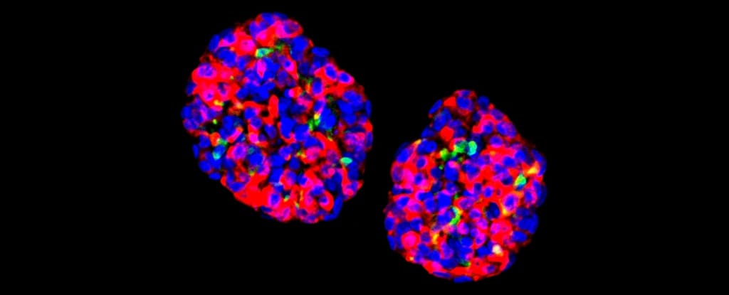
Harnessing this intrinsic developmental proximity, the Weill Cornell team successfully isolated adult gastric stem cells from human stomach biopsies obtained via routine, outpatient upper endoscopy. Once placed in specialized 3D culture matrices enriched with key growth factors (including Wnt3a, R-spondin, and epidermal growth factor), these gastric stem cells rapidly multiplied into self-organizing three-dimensional mini-tissues known as gastric organoids [1].
The scientists then introduced a transient molecular differentiation protocol. Over a span of ten to twelve days, the gastric organoids were systematically exposed to specific signaling cues that silenced the stomach mucosal gene program and activated endocrine fate specification. The resulting micro-clusters underwent a dramatic phenotypic metamorphosis: they stopped producing stomach mucin and gastric enzymes, and instead began expressing insulin, C-peptide (a clinical marker of endogenous insulin synthesis), and the vesicular packaging machinery necessary for endocrine secretion [1] [5].
These engineered structures were designated GINS (Gastric Insulin-Secreting) cells. Electron microscopy of mature GINS organoids revealed dense-core neuroendocrine secretory granules morphologically indistinguishable from natural pancreatic beta cells. When challenged with varying glucose concentrations in culture, GINS cells demonstrated genuine physiological glucose responsiveness: releasing minimal baseline insulin at low glucose levels (under 3 millimolar) and surging insulin production when exposed to high glucose concentrations (above 15 to 20 millimolar) [1].
Transcription Factor Reprogramming: The Role of PDX1, NGN3, and MAFA
At the molecular core of this cellular alchemy lies a triad of master transcriptional regulators known in developmental biology as the ‘pioneer factors’: PDX1 (Pancreatic and Duodenal Homeobox 1), NGN3 (Neurogenin 3), and MAFA (MAF BZIP Transcription Factor A) [2] [4].
- PDX1: Functions as the foundational master architect of pancreatic identity, binding closed chromatin regions to unmask beta-cell-specific promoters and activating insulin gene transcription.
- NGN3: Serves as the transient developmental switch that directs endodermal progenitor cells to abandon the intestinal epithelial fate and commit irreversibly to the endocrine lineage.
- MAFA: Acts as the terminal maturation factor, binding the C1/RIPE3b enhancer element inside the insulin promoter to establish glucose-responsive insulin exocytosis and adult beta-cell stability.
In the Weill Cornell protocol, transient activation of these transcription factors, combined with inhibition of notch and TGF-beta signaling, proved sufficient to orchestrate the complete epigenetic rewiring of gastric antrum progenitors. The cells permanently downregulated stomach-specific markers such as mucin 5AC (MUC5AC) while stably activating the insulin gene along with essential endocrine machinery, including prohormone convertase 1/3 (PCSK1) and the sulfonylurea receptor (SUR1/KCNJ11 potassium channel complex) [1] [2].
Preclinical Evidence: Reversing Hyperglycemia in Animal Models
The definitive test of any cellular therapy lies in its therapeutic efficacy in living physiology. To evaluate whether GINS cells could rescue severe diabetic hyperglycemia, researchers transplanted human GINS organoids under the renal capsules and into the abdominal cavity of immunocompromised diabetic mice whose native beta cells had been chemically eradicated with streptozotocin [1].
The physiological results were extraordinary. Within ten to fourteen days post-implantation, the transplanted GINS organoids successfully vascularized, integrating into the host blood supply. Blood glucose concentrations in the diabetic mice, which had soared above 450 mg/dL, rapidly plummeted back into the normal physiological range (90 to 120 mg/dL) [1].
Crucially, the GINS grafts maintained sustained glycemic control for more than six months without a single episode of reactive hypoglycemia. When researchers performed oral glucose tolerance tests, the transplanted animals cleared glucose with kinetics nearly identical to healthy non-diabetic controls. Furthermore, when the GINS organoid grafts were surgically removed after six months, the mice immediately relapsed into severe diabetic hyperglycemia, conclusively proving that the reprogrammed human stomach cells were entirely responsible for maintaining metabolic survival [1].
Circumventing Immune Rejection: Autologous Transplants and Microencapsulation
The Achilles’ heel of conventional organ and tissue transplantation has always been allogeneic immune rejection. Because cadaveric donor organs possess foreign human leukocyte antigens (HLA), the recipient’s immune system launches an aggressive rejection response unless paralyzed by heavy immunosuppressive drugs like tacrolimus, cyclosporine, and mycophenolate mofetil [3] [4].
Regenerative Comparison: Beta Cell Replacement Strategies
Cadaveric Islet Allografts
Cell Source: Deceased organ donors; severely limited supply.
Immunology: Foreign HLA profiles; requires lifelong toxic immunosuppressive medications.
Viability: High graft attrition rate over 3 to 5 years.
Clinical Status: Approved for extreme, life-threatening hypoglycemic unawareness.
Pluripotent Stem Cells (iPSC)
Cell Source: Induced pluripotent or embryonic cell lines.
Immunology: Allogeneic lines require immunosuppression or CRISPR immune-cloaking edits.
Viability: Months-long multi-stage manufacturing with teratoma risks.
Clinical Status: Active Phase 1/2 clinical trials (e.g., Vertex VX-880).
Reprogrammed Gastric (GINS)
Cell Source: Autologous adult gastric stem cells from routine patient endoscopy.
Immunology: 100% genetically identical to patient; zero foreign allo-rejection risk.
Viability: Rapid 10-day organoid conversion with robust physiological glucose sensing.
Clinical Status: Preclinical stage progressing toward human clinical translation.
GINS technology fundamentally solves the donor-matching and allo-rejection dilemmas. Because the starting cells are harvested directly from the patient via a routine, ten-minute gastroscopy biopsy, the resulting GINS cells carry the patient’s own native genome. When reintroduced into the patient, the immune system recognizes them as ‘self’. However, for type 1 diabetes patients whose immune systems destroyed their original beta cells, one critical challenge remains: preventing the underlying autoimmunity from attacking the newly implanted GINS cells. Researchers are addressing this by placing GINS organoids inside semi-permeable biocompatible hydrogel microcapsules. These microcapsules allow oxygen, nutrients, and insulin to diffuse freely while physically blocking large T-cell antibodies and cytotoxic lymphocytes from touching the cells [1] [3].
The Future of Diabetes Therapeutics: From Daily Injections to Living Cellular Cures
The transition from animal models to human clinical trials represents the next major milestone. Biotech spin-offs and academic consortia are currently optimizing automated, closed-loop bioreactors capable of manufacturing billions of clinical-grade GINS cells under Good Manufacturing Practice (GMP) conditions. Early clinical pipelines envision implanting these micro-encapsulated cellular units into the highly vascularized subcutaneous space or the omental pouch of diabetic individuals [1] [4].
Once implanted, these living cellular devices could function as an autonomous internal pancreas for years at a time, liberating patients from finger pricks, carbohydrate calculations, and insulin injections. Beyond type 1 diabetes, this technology offers an essential therapeutic reservoir for hundreds of millions of advanced type 2 diabetes patients whose pancreatic reserves have completely collapsed after decades of metabolic exhaustion [2] [5].
The discovery that human stomach cells can be converted into insulin-producing bio-factories stands as a testament to the astonishing plasticity of human biology. By looking inward to our own tissues, regenerative medicine is transforming diabetes from a lifelong chronic sentence into a biologically curable condition [1] [3].
Frequently Asked Questions
Common reader questions about this study
Why are stomach cells chosen for insulin-producing cellular reprogramming?
During early embryonic development, the stomach and pancreas arise from adjacent regions of the anterior foregut endoderm. Gastric stem cells located in the stomach antrum retain epigenetic memory closely aligned with pancreatic progenitors, making them vastly easier to transdifferentiate into functional insulin-producing cells than skin or blood cells.
What are GINS cells and how do they function?
GINS stands for Gastric Insulin-Secreting cells. These are laboratory-engineered micro-tissues created by converting adult gastric stem cells into glucose-responsive endocrine cells equipped with insulin granules and molecular glucose sensors like GLUT2 and glucokinase.
Can GINS cell therapy cure both type 1 and type 2 diabetes?
GINS therapy holds primary therapeutic promise for type 1 diabetes, where autoimmune destruction eliminates native pancreatic beta cells. However, it also offers significant benefit for advanced type 2 diabetes patients suffering from severe beta-cell exhaustion and chronic insulin deficiency.
Does receiving GINS cells require lifelong immunosuppressive drugs?
No, because GINS cells are derived autologously from the patient's own stomach mucosa through a standard, minimally invasive gastroscopy. Because the transplanted cells share the patient's identical genetic signature, the host immune system recognizes them as 'self', avoiding organ rejection.
References
- ↩ Nature Cell Biology: Conversion of adult human gastric stem cells into insulin-secreting cells protects against diabetes
- ↩ Cell Stem Cell: In vivo reprogramming of adult pancreatic and gastric cells into insulin-producing beta cells
- ↩ The New England Journal of Medicine: Stem-Cell-Derived Islet Replacement Therapy for Type 1 Diabetes
- ↩ Nature Reviews Endocrinology: Cellular plasticity and transdifferentiation in diabetes therapy
- ↩ Diabetes: Generation of functional insulin-producing cells from human gastric endoderm

No comments yet. Be the first to join the conversation.