


Key Findings
- Sustained weight loss of 5% reduces hepatic steatosis, while 7% to 10% loss resolves steatohepatitis and helps regress early fibrosis.
- Primary-care FIB-4 scores combine age, AST, ALT, and platelets to detect silent scarring even when liver enzymes look normal.
- Activated hepatic stellate cells transform silent fat buildup into dense collagen scars, driving portal hypertension and cirrhosis.
Few human organs absorb silent metabolic injury as long as the liver. Internal liver tissue holds very few sensory pain fibers [2]. Fat droplets and microscopic collagen scars can therefore accumulate for ten to twenty years before jaundice or abdominal swelling appears [1, 2].
Global hepatology societies recently updated fatty liver terminology from nonalcoholic fatty liver disease (NAFLD) to metabolic dysfunction-associated steatotic liver disease (MASLD) to spotlight its true drivers [1]. Insulin resistance, central adiposity, regular alcohol intake, and chronic viral hepatitis now drive most progressive scarring worldwide [1, 2].
Clinical guidelines from AASLD, EASL, and The Lancet trace the exact biological bridge separating reversible hepatic fat accumulation from irreversible decompensated cirrhosis [1, 2, 3]. Catching elevated liver risk early overlaps directly with monitoring subtle early signs of prediabetes and checking everyday habits covered in moderate alcohol and organ risk science.
Why This Matters: Catching Silent Fibrosis Before Decompensation
Most people assume liver disease triggers sharp upper-right rib pain early on. In clinical reality, compensated cirrhosis often presents with completely normal daily energy and unremarkable routine blood tests until portal blood pressure rises sharply [2, 3]. Because liver cells carry huge spare reserve capacity, silent injury marches forward quietly for years [2].
Once cirrhosis crosses into decompensation, marked by abdominal fluid (ascites), esophageal bleeding, or confused mental alertness (hepatic encephalopathy), short-term survival drops dramatically without specialist care or liver transplantation [2, 3]. Globally, cirrhosis accounts for over one million annual deaths across alcohol-related disease, viral hepatitis, and metabolic steatosis [2].
Preventing that irreversible cliff begins years earlier inside routine primary care visits. Non-invasive blood indices and elastography stiffness scans catch advancing liver fibrosis cleanly long before symptoms emerge [1, 2]. Addressing dietary triggers explored in our report on harmful daily eating habits reduces liver fat pressure directly. Early awareness turns progressive organ scarring into a manageable, largely preventable metabolic condition [1, 2].
How the Study Was Done: AASLD, Lancet, and EASL Consensus
Modern liver damage management rests on three major clinical evidence syntheses [1, 2, 3]. First, Mary Rinella and co-authors published updated American Association for the Study of Liver Diseases (AASLD) practice guidance in Hepatology in 2023 [1]. That guidance defined risk-stratification pathways using FIB-4 blood ratios and vibration-controlled transient elastography to spare patients needless liver biopsies [1].
Second, Pere Ginès and international hepatologists authored a comprehensive medical seminar on liver cirrhosis in The Lancet in 2021 [2]. They integrated worldwide epidemiology, hepatic stellate cell biology, and clinical criteria dividing stable compensated scarring from acute decompensation [2].
Third, Paolo Angeli and the European Association for the Study of the Liver (EASL) published clinical practice guidelines in the Journal of Hepatology in 2018 [3]. Their protocols guide bedside management of ascites, variceal bleeding, and bacterial infections inside decompensated cirrhosis clinics [3].
How Fat Turns Into Scar Tissue Inside Hepatocytes
Healthy hepatocytes work nonstop as the body’s chemical processing hub [2]. They filter absorbed sugars, build clotting proteins, and clear gut toxins from incoming portal venous blood [2]. Chronic excess dietary sugar, fructose, saturated fats, and visceral insulin resistance overwhelm hepatocyte lipid export capacity [1].
Triglyceride droplets begin accumulating inside liver cells, producing hepatic steatosis [1, 2]. When excess fat occupies more than five percent of hepatocyte volume, medical guidelines classify the liver as steatotic [1]. At this early stage, liver architecture remains intact and fully reversible if metabolic stress lifts [1]. Simple dietary cleanup and weight reduction rapidly drain excess fat droplets before fibrotic scarring takes hold [1].
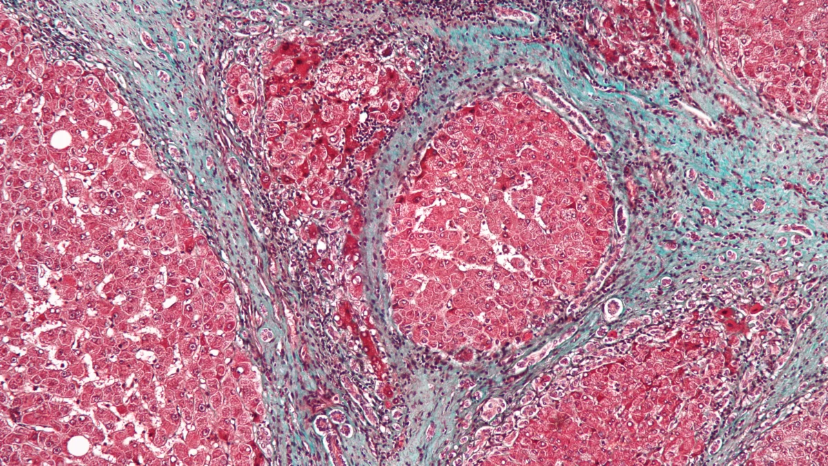
However, oxidized lipid byproducts and gut bacterial endotoxins irritate resident liver immune macrophages called Kupffer cells [2]. Inflammatory signaling activates dormant hepatic stellate cells tucked beside hepatic sinusoids [2]. Activated stellate cells morph into scar-weaving myofibroblasts, pumping thick collagen fibers into surrounding tissue spaces and gradually stiffening soft liver tissue into cirrhotic nodules [1, 2].
How Chronic Liver Injury Progresses Toward Cirrhosis
- Stage 4
Cirrhosis and Portal Hypertension
Compensated vs Decompensated Cirrhosis: The Critical Clinical Split
Not everyone diagnosed with early fibrosis progresses to liver failure, and cirrhosis itself behaves like two vastly different diseases across stages [2].
During compensated cirrhosis, remaining healthy hepatocytes work overtime to maintain clotting factors, albumin synthesis, and bilirubin clearance despite dense microscopic scars [2]. Patients frequently feel normal while pressure quietly climbs inside the portal vein leading from intestines to liver [2, 3]. Regular six-month ultrasound surveillance still matters at this stable stage because stiff, regenerating nodules carry higher risk for hepatocellular carcinoma [1, 2].
When portal hypertension crosses critical thresholds alongside declining liver cell mass, patients enter decompensated cirrhosis [2, 3]. Blood backs up into delicate esophageal veins that can rupture dangerously, fluid leaks into the abdominal cavity (ascites), and unmetabolized ammonia reaches the brain causing hepatic encephalopathy [2, 3]. EASL protocols target reversing triggers like hidden infections or bleeding immediately because decompensated complications escalate rapidly [3]. Treating acute bacterial peritonitis early and managing sodium fluid retention extends hospital-free survival substantially [3].
Table 1 summarizes biological drivers, clinical warning signs, and non-invasive monitoring stages across liver injury progression [1, 2, 3].
| Clinical Stage | Dominant Cellular Mechanism | Common Symptoms | Evidence-Based Intervention |
|---|---|---|---|
| Simple Steatosis (MASLD) [1] | Reversible triglyceride droplet buildup | Usually silent; mild enzyme shifts possible | Calorie balance, resistance exercise, blood sugar control |
| Steatohepatitis & Fibrosis [1, 2] | Kupffer inflammation and stellate collagen bands | Silent or subtle fatigue; tracked via FIB-4 & ultrasound | Structured weight loss, metabolic therapy, complete alcohol avoidance |
| Compensated Cirrhosis [2] | Regenerative nodules raising portal pressure | Often symptom-free despite structural scarring | Ultrasound cancer surveillance, beta-blockers if indicated |
| Decompensated Cirrhosis [2, 3] | Portal hypertension & hepatocyte failure | Ascites fluid, variceal bleeding, brain fog, jaundice | Specialist diuretics, endoscopic banding, transplant evaluation |
Compiled from Rinella et al. (2023 AASLD), Gines et al. (2021 Lancet), and EASL Guidelines (2018).
Non-Invasive Screening: FIB-4 and Liver Stiffness Scans
Doctors no longer wait for an invasive needle biopsy just to check whether metabolic fatty liver has begun scarring [1]. AASLD guidance recommends starting with the simple FIB-4 calculation built from routine blood counts and liver enzymes: patient age, AST, ALT, and platelet count [1].
Why do blood platelets matter for liver staging? As advancing liver fibrosis increases resistance inside portal blood channels, the spleen traps platelets out of circulation, causing circulating platelet numbers to drift downward subtly over years [1, 2].
People whose FIB-4 score lands in an intermediate or elevated zone next undergo transient elastography, a painless 5-minute ultrasound pulse measuring acoustic wave velocity through liver tissue [1]. Faster sound-wave propagation indicates stiffer fibrotic liver parenchyma accurately without touching needles [1]. Low-risk FIB-4 scores allow primary doctors to avoid unnecessary biopsies while focusing repeat monitoring on metabolic control [1]. High stiffness measurements promptly route patients to gastroenterology specialists for targeted endoscopic screening and antifibrotic counseling [1, 2]. Understanding routine lab markers connects closely with broader tracking insights in our guide on interpreting clinical blood biomarkers.
Reversing Early Damage Through Diet, Weight Loss, and Abstinence
Because hepatocyte regeneration outpaces collagen accumulation during early stages, sustained weight loss and alcohol cessation deliver dramatic biological recovery [1, 2]. AASLD trials confirm losing roughly 5 percent of body weight trims hepatic steatosis measurably, while 7 to 10 percent weight reduction resolves inflammatory steatohepatitis and helps regress early fibrosis [1]. Adding brisk aerobic exercise plus twice-weekly resistance sessions improves liver fat independently of scale weight changes by pulling circulating glucose directly into active skeletal muscle [1].
Cutting liquid sugar beverages rich in free fructose removes a potent direct driver of liver fat synthesis [1]. Replacing refined pastries and sugary sodas with whole fiber-rich foods, plain water, and unsweetened coffee lowers metabolic fat delivery into hepatic portal blood [1]. Meanwhile, for alcohol-associated steatosis or mixed metabolic injury, total abstinence offers the strongest lever halting stellate cell scar production, even in early compensated cirrhosis [2]. Vaccinating against hepatitis A and hepatitis B further prevents sudden acute viral hits atop an already stressed liver [1, 2].
What the study cannot tell us: Limits of Blood Enzymes
Patients frequently assume normal AST and ALT values on an annual blood panel guarantee a healthy, unscarred liver [1]. In clinical reality, many adults carrying advanced stage F3 fibrosis or silent cirrhosis still show completely normal routine transaminase enzymes [1, 2]. Relying solely on AST/ALT misses silent disease unless age and platelets factor into FIB-4 calculations [1].
Genetic variation, such as PNPLA3 gene variants, also alters why one person with mild overweight develops scarring quickly while another avoids fibrosis for decades [1, 2]. Unregulated herbal “liver detox” supplements and weight-loss teas also remain a leading cause of unexpected acute drug-induced liver injury worldwide [2]. Clinical trials likewise continue testing newer metabolic and thyroid-beta drugs targeting fibrotic MASH pathways alongside lifestyle care [1].
Adults living with type 2 diabetes, central obesity, or regular alcohol exposure benefit from asking a primary doctor for FIB-4 screening [1, 2]. Early non-invasive checks catch reversible fatty liver years before visible jaundice or emergency swelling appears [1, 2].
Frequently Asked Questions About Liver Steatosis and Cirrhosis
Frequently Asked Questions
Can fatty liver disease genuinely reverse without prescription pills?
Yes, in early stages. AASLD clinical guidance shows losing 5 percent of body weight measurably clears liver fat, while 7 to 10 percent sustained weight loss resolves inflammation and can regress early fibrosis.
Why did medical guidelines rename NAFLD to MASLD?
Experts adopted metabolic dysfunction-associated steatotic liver disease (MASLD) to replace exclusionary ‘nonalcoholic’ language with an accurate diagnosis rooted in cardiometabolic risk like insulin resistance and diabetes.
If my AST and ALT liver enzyme blood tests are normal, am I safe from cirrhosis?
Not necessarily. Many patients with advanced liver fibrosis or compensated cirrhosis retain normal AST and ALT enzymes, which is why doctors compute composite indices like FIB-4 and use elastography scans.
Do herbal liver cleanse drinks repair fatty liver faster?
No. Clinical guidelines note unregulated botanical ‘detox’ extracts can actually trigger acute drug-induced hepatotoxicity, whereas balanced calorie reduction, aerobic exercise, and stopping alcohol genuinely reduce liver fat.
Technical Terms and Definitions
Scientific terms used in this article
- MASLD (formerly NAFLD)Metabolic Fatty Liver Disease
- Fat accumulation exceeding liver thresholds linked directly to insulin resistance, obesity, hypertension, or type 2 diabetes.
- Hepatic Stellate CellsLiver Scar-Forming Cells
- Specialized perivascular liver cells that turn into collagen-depositing myofibroblasts during chronic inflammation.
- Portal HypertensionHigh Liver Vein Pressure
- Elevated blood pressure inside the portal venous system caused by rigid cirrhotic nodules blocking blood flow.
- FIB-4 IndexNon-Invasive Fibrosis Score
- A validated screening ratio combining patient age, AST, ALT, and platelet count to spot advanced liver scarring.
Scientific References and Primary Literature
AASLD Practice Guidance on the clinical assessment and management of nonalcoholic fatty liver disease
- Journal
- Hepatology
- Study Type
- Report
- Year
- 2023
DOI10.1097/HEP.0000000000000323
Timeline
- 2018
EASL Decompensated Care Rules
European hepatology guidelines standardize emergency management of ascites, variceal bleeds, and encephalopathy.
- 2021
Lancet Global Cirrhosis Seminar
Synthesized worldwide transition criteria dividing compensated stability from portal hypertension failure.
- 2023
AASLD MASLD Screening Guidance
Established primary-care FIB-4 + elastography workflows and 5% to 10% weight-loss fibrosis regression goals.
References
- ↩ Rinella, M. E., Neuschwander-Tetri, B. A., Siddiqui, M. S., et al. (2023). AASLD Practice Guidance on the clinical assessment and management of nonalcoholic fatty liver disease. Hepatology 77(5), 1797-1835.
- ↩ Gines, P., Krag, A., Abraldes, J. G., Sola, E., Fabrellas, N., & Kamath, P. S. (2021). Liver cirrhosis. The Lancet 398(10308), 1359-1376.
- ↩ Angeli, P., Bernardi, M., Villanueva, C., et al. (2018). EASL Clinical Practice Guidelines for the management of patients with decompensated cirrhosis. Journal of Hepatology 69(2), 406-460.



No comments yet. Be the first to join the conversation.