


Key Findings
- A breast screening AI cut missed cancers by 9.4% (US) and 2.7% (UK) while cutting false alarms by 5.7% and 1.2% [1].
- Deep retinal fundus networks reached 0.991 AUC on EyePACS-1 and 0.990 AUC on Messidor-2 for diabetic retinopathy triage [2].
- Dataset shift, demographic under-representation, and automation bias require prospective hospital trials alongside human doctors [3].
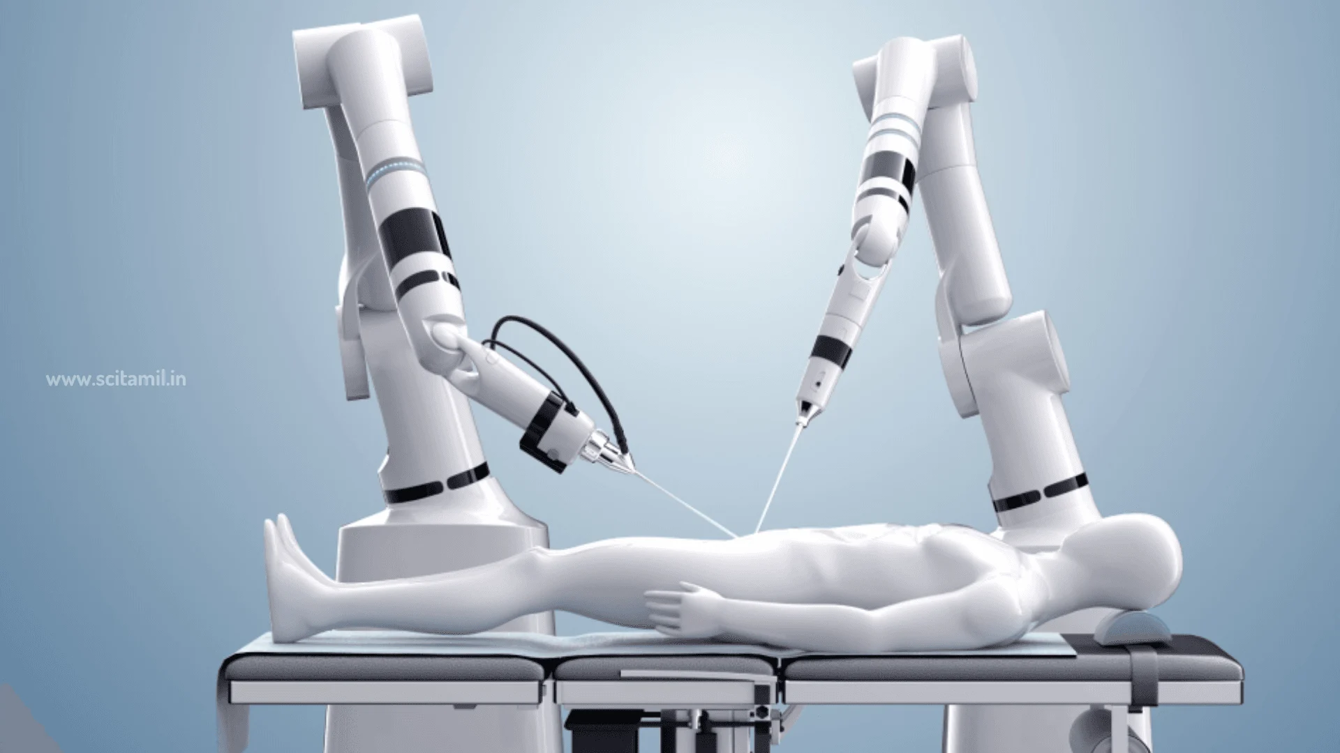
Modern hospitals generate thousands of mammograms, retinal photographs, CT slices, and pathology slides faster than specialists can review them. Deep learning computer vision now steps into that bottleneck as a tireless second reader [1, 2, 3].
In landmark clinical validations across tens of thousands of patients, convolutional neural networks caught diabetic eye disease with area-under-curve scores above 0.99 [2]. Across US mammography scans, deep learning also lowered missed breast cancers by 9.4 percentage points [1].
High-performance medical computing works best when paired with trained doctors rather than replacing them [3]. For direct head-to-head diagnostic trial comparisons, read our deep dive on whether AI diagnoses illness as accurately as doctors alongside our guide on spotting early metabolic complications before diabetes progresses.
Why This Matters: Visual Fatigue and Screening Access
Missed early lesions and false alarms both carry steep human costs. Reviewing hundreds of normal screening scans daily induces perceptual fatigue in busy radiology departments, raising error odds near shift ends [1, 3]. Subtle hairline tumor margins or tiny retinal hemorrhages can blur past tired eyes after dozens of consecutive patient screens [1, 2].
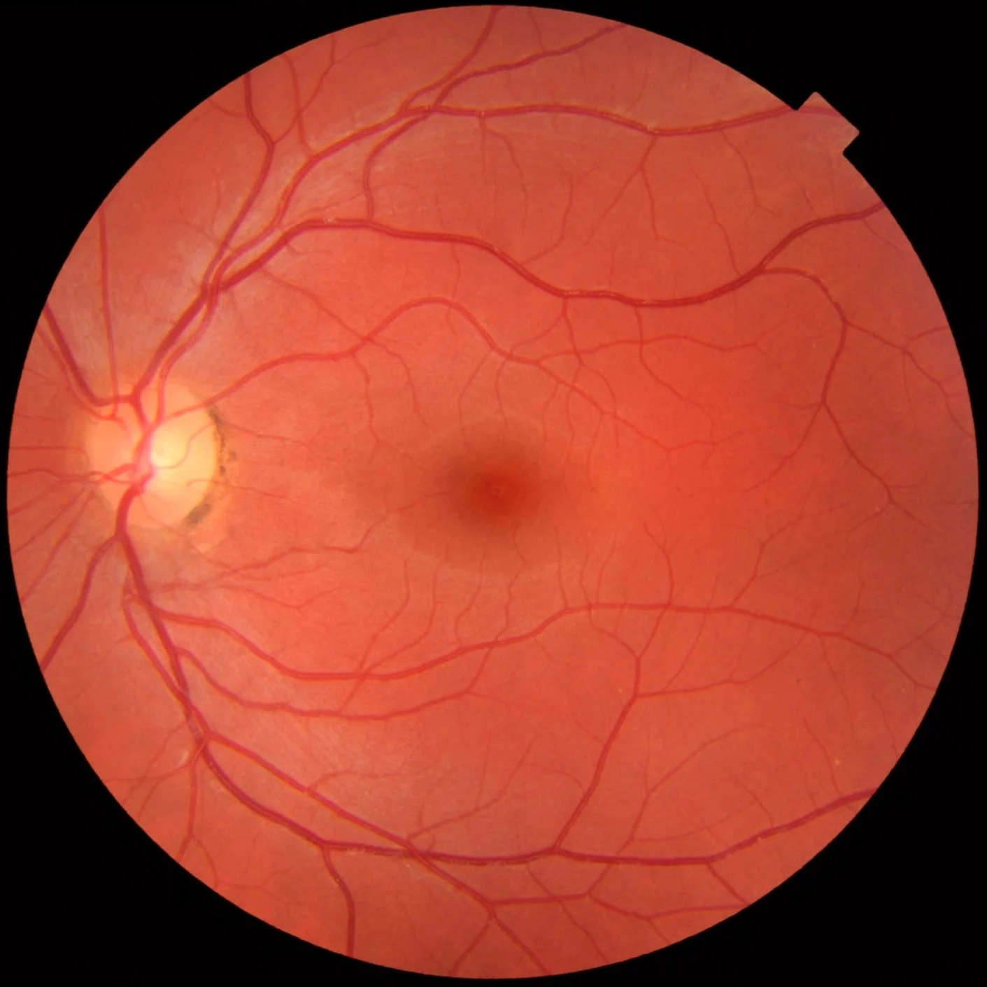
Meanwhile, rural clinics worldwide lack enough ophthalmologists to inspect retinal photos for hundreds of millions of adults living with diabetes [2, 3]. Automated retinal triage software flags abnormal images instantly in primary clinics so specialists treat high-risk eyes before vision fades permanently [2, 3]. Rapid automated triage keeps routine normal cases moving while steering specialist hours directly toward patients who need immediate biopsy or laser care [2, 3].
Bridging digital sensor data and patient care builds on pattern-recognition architectures also shaping hardware systems explored in our report on autonomous decision systems in robotics.
How the Study Was Done: Multi-Site Reader Trials
Rigorous medical AI evaluation starts far beyond accuracy claims on clean benchmark toys. Researchers test algorithms against independent hospital populations, unseen scanners, and live human specialist panels [1, 2, 3].
In JAMA in 2016, Varun Gulshan and colleagues trained a deep convolutional neural network on 128,175 retinal photographs graded repeatedly by ophthalmologists [2]. They then evaluated performance on two completely separate external datasets: EyePACS-1 (9,963 images) and Messidor-2 (1,748 images) [2].
In Nature in 2020, Scott McKinney, Christopher Kelly, and international collaborators evaluated an AI mammography model across large UK and US screening cohorts [1]. They also ran a direct reader study pitting the algorithm against six board-certified US radiologists on identical screening mammograms [1].
Eric Topol synthesized those imaging breakthroughs alongside electronic health records and clinical workflow trials in Nature Medicine in 2019, highlighting both clinical gains and real-world deployment hurdles [3].
How Computer Vision Reads Mammograms and Retinas
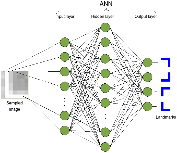
Unlike rule-based software written with rigid checklists, deep convolutional networks learn hierarchical pixel statistics directly from labeled medical archives [2, 3]. Early network layers detect faint edges, micro-calcifications, or hairline capillary bulges [1, 2].
Deeper layers combine subtle spatial textures across entire organs, outputting calibrated risk scores rather than simple yes-no tags [1, 2]. Tuning operating thresholds lets clinics favor either maximum sensitivity, catching almost every possible lesion, or high specificity to spare healthy people unnecessary anxiety [2].
How Medical Imaging AI Assists Screening Workflows
In Gulshan and colleagues’ retinal study, choosing a high-sensitivity cut-point delivered 97.5 percent sensitivity on EyePACS-1 and 96.1 percent on Messidor-2 [2]. At that same operating setting, specificity held strong at 93.4 percent on EyePACS-1 and 93.9 percent on Messidor-2 [2].
Shifting the decision cut-point toward high specificity raised specificity to 98.1 percent on EyePACS-1 and 98.5 percent on Messidor-2, while sensitivity remained 90.3 percent and 87.0 percent respectively [2]. Clinics tune those thresholds according to local specialist capacity [2, 3]. Remote screening vans prioritize high sensitivity so no threatened eye slips past. Meanwhile, packed hospital eye clinics lean toward high specificity to keep surgical backlogs manageable [2, 3].
Mammography Trials: Fewer Missed Cancers and Recalls
Screening mammography poses a notoriously delicate balance between catching tiny tumors early and avoiding false biopsy scares [1]. In the United Kingdom, national screening relies on two independent radiologists reading every mammogram, consuming scarce specialist hours [1]. Finding subtle early breast calcifications requires deep visual concentration across thousands of nearly identical normal breast scans [1].
McKinney and colleagues found their ensemble AI system cut false-positive rates by 5.7 percent in US scans and 1.2 percent in UK scans [1]. Simultaneously, false-negative rates dropped by 9.4 percent in US cases and 2.7 percent in UK evaluations [1]. Why did margins differ between the two countries? In the US, single radiologists read screening exams more conservatively, leaving broader room to trim false-positive biopsy callbacks [1].
Even more impressively, simulating an automated second-reader setup reduced second-reader radiologist workload dramatically while maintaining non-inferior accuracy across UK clinical standards [1]. Under that collaborative triage setup, the system only flagged discordant cases between human and machine for a third human arbiter [1]. Table 1 summarizes validated screening benchmarks across ophthalmology and breast imaging trials [1, 2, 3].
| Clinical Domain | Study & Year | Validation Dataset | Verified Diagnostic Benchmark |
|---|---|---|---|
| Breast Cancer Screening [1] | McKinney et al. 2020 (Nature) | US & UK screening mammograms | -5.7% / -1.2% false positives; -9.4% / -2.7% false negatives |
| Diabetic Retinopathy [2] | Gulshan et al. 2016 (JAMA) | EyePACS-1 (n=9,963) & Messidor-2 | 0.991 & 0.990 AUC; 96.1% to 97.5% sensitivity |
| Human-AI Workflow [3] | Topol 2019 (Nature Medicine) | Cross-specialty synthesis | Faster triage, reduced keyboard clerical burden, safety oversight |
Compiled from McKinney et al. (2020), Gulshan et al. (2016), and Topol (2019).
Beyond Imaging: Triage, Records, and Clinician Burnout
Radiology and eye clinics represent only the most mature layer of clinical software [3]. As Eric Topol observed, hospital doctors frequently spend nearly as many hours typing administrative electronic chart notes as speaking directly with patients [3].
Ambient clinical voice transcription and structured medical summarization aim to lift keyboard workloads so clinicians restore eye contact at the bedside [3]. Speech-recognition infrastructure shares acoustic processing roots with tools examined in our guide on modern neural speech synthesis models.
Emergency departments also deploy machine-learning triage alerts tracking vital signs, blood lactate trends, and continuous bedside waveforms to spot silent deterioration early [3]. In digital pathology, whole-slide scanners digitize lymph node biopsies at high magnification so algorithms flag microscopic tumor clusters that might hide inside massive glass slides [3].
Dermatology and primary care clinics also test image classifiers trained on thousands of pigmented skin moles to flag possible melanomas for faster biopsy referral [3]. Cardiology units similarly apply neural classifiers to standard 12-lead ECG traces to identify asymptomatic left-ventricular dysfunction before heart failure symptoms erupt [3].
When designed cleanly, predictive alerts buy bedside nurses precious minutes rather than flooding screens with noisy false alarms [3]. Hospital teams gain real clinical value only when software plugs smoothly into existing radiology workstations and electronic health records without extra mouse clicks [3]. Clear user interfaces also display calibrated uncertainty intervals rather than brittle confidence labels [3].
Compare algorithmic screening directly with human specialists in our review of AI vs doctors diagnostic accuracy trials. Evidence-based clinical workflow design also shapes outpatient protocols explored in our report on pediatric dermatology management.
Privacy architecture matters equally whenever medical algorithms train across sensitive patient files [3]. Federated learning techniques keep raw patient X-rays inside each hospital’s locked servers, sharing only anonymized mathematical weight updates across research consortia [3]. That decentralized workflow enables rare-disease collaboration across countries while respecting strict medical confidentiality standards [3].
What the study cannot tell us: Bias, Drift, and Safety Gaps
Despite glittering retrospective AUC figures, clinical AI faces four stubborn hazards inside real hospitals [1, 3]. First, dataset shift happens whenever software trained on one scanner brand or wealthy urban hospital encounters rural clinics using older equipment [3].
Second, demographic bias creeps in quietly when skin conditions, chest anatomy, or genetic disease burdens are under-represented during training [3]. Models trained primarily on fair skin tones, for example, can drop noticeably when evaluated on darker skin dermatology photos [3]. Auditing error rates separately across age groups, sexes, and ancestries prevents hidden disparities before national rollout [3].
Third, automation bias tempts tired clinicians to agree passively with confident-looking computer scores even when subtle clinical clues contradict the algorithm [3]. Medical educators increasingly train resident doctors to treat model saliency heatmaps skeptically and cross-check raw scan slices independently [3].
Fourth, retrospective accuracy on stored image archives never automatically proves improved patient survival [3]. Regulators such as the US Food and Drug Administration increasingly examine real-world post-market drift alongside pre-approval validation studies [3]. Topol emphasizes that prospective randomized clinical trials inside active clinics remain essential before granting autonomous systems unchecked clinical authority [3]. Safe healthcare software must treat algorithms as transparent decision-support tools accountable to human medical judgment [3].
Frequently Asked Questions About AI in Medicine
Frequently Asked Questions
Can medical imaging AI replace radiologists or eye doctors completely?
No. Clinical leaders emphasize human-AI partnership. Software excels at tireless pixel screening and workflow prioritization, whereas physicians integrate ambiguous patient histories, complex comorbidities, ethics, and physical exams.
How accurately does deep learning detect diabetic retinopathy?
In a 2016 JAMA validation study across two external datasets, a deep convolutional network achieved areas under the ROC curve of 0.990 and 0.991, reaching 96 to 97.5 percent sensitivity for referable eye disease.
Did AI reduce errors on mammogram screening scans?
Yes. In a 2020 Nature evaluation across UK and US cohorts, the AI model achieved absolute drops of 5.7 percent (US) and 1.2 percent (UK) in false positives alongside 9.4 percent (US) and 2.7 percent (UK) reductions in missed cancers.
Why can medical AI stumble outside the hospital where it was trained?
Different imaging hardware calibration, patient demographics, and disease prevalence, known as dataset shift, can degrade unverified models, making local hospital validation mandatory.
Technical Terms and Definitions
Scientific terms used in this article
- Area Under the ROC Curve (AUC)Overall Classifier Accuracy Score
- A metric from 0.5 to 1.0 summarizing how cleanly an algorithm separates disease cases from healthy scans across thresholds.
- Sensitivity and SpecificityDetection vs False-Alarm Rates
- Sensitivity measures true disease caught; specificity measures healthy cases correctly cleared without false alarms.
- Convolutional Neural Network (CNN)Visual Pattern Deep Learning
- A layered neural architecture built specifically to detect edges, shapes, and anomalies across two-dimensional medical images.
- Dataset ShiftHospital Generalization Drift
- Accuracy loss occurring when a model trained on one hospital or scanner faces unfamiliar real-world equipment or populations.
Scientific References and Primary Literature
International evaluation of an AI system for breast cancer screening
- Journal
- Nature
- Study Type
- Cohort
- Year
- 2020
DOI10.1038/s41586-019-1799-6
Timeline
- 2016
Retinal Deep Learning Validated
Gulshan et al. demonstrated 0.99+ AUC screening diabetic eye disease across external cohorts.
- 2019
Clinical Convergence Mapped
Topol outlined human-AI workflows, automation bias, and EHR voice transcription horizons.
- 2020
Mammography AI Reader Study
McKinney et al. cut missed breast cancers by 9.4% (US) and false alarms by 5.7% (US).
References
- ↩ McKinney, S. M., Sieniek, M., Godbole, V., et al. (2020). International evaluation of an AI system for breast cancer screening. Nature 577(7788), 89-94.
- ↩ Gulshan, V., Peng, L., Coram, M., et al. (2016). Development and Validation of a Deep Learning Algorithm for Detection of Diabetic Retinopathy in Retinal Fundus Photographs. JAMA 316(22), 2402-2410.
- ↩ Topol, E. J. (2019). High-performance medicine: the convergence of human and artificial intelligence. Nature Medicine 25(1), 44-56.



No comments yet. Be the first to join the conversation.